Colorectal cancer peritoneal metastasis model and establishment method thereof

A metastasis model, colorectal cancer technology, applied in botany equipment and methods, biochemical equipment and methods, pharmaceutical formulations, etc., can solve the problems of tumor inoculation failure, many complications, and long preparation cycle

Inactive Publication Date: 2016-10-26
FUDAN UNIV SHANGHAI CANCER CENT
View PDF1 Cites 3 Cited by
  • Summary
  • Abstract
  • Description
  • Claims
  • Application Information

AI Technical Summary

Problems solved by technology

Colon cancer mouse models in the prior art either have a long preparation period and poor tumor uniformity; or fail to fully simulate the natural metastasis process and have many complications; or have complicated preparation operations, low success rate, and unstable models
Due to the thin and slender intestinal wall of mice, the injection needle can easily pierce the intestinal wall, which makes in situ injection of the colon wall difficult, or the failure of tumor inoculation due to the puncture of the intestinal wall, or the leakage of fluid in many places in the peritoneal cavity outside the intestine. to plant
Even if the tumor is successfully induced, the tumor tissue at the inoculation site grows too large, causing intestinal obstruction and death of the mice

Method used


Image

  • Colorectal cancer peritoneal metastasis model and establishment method thereof
  • Colorectal cancer peritoneal metastasis model and establishment method thereof
  • Colorectal cancer peritoneal metastasis model and establishment method thereof

Examples


Embodiment 1

[0038] Example 1 Establishment of peritoneal metastasis model by intraperitoneal injection of colorectal cancer cell lines

[0039] 1. Animal model

[0040] (1) intraperitoneal injection, 2*10 5 cells / only

[0041] 2. In vivo fluorescence imaging

[0042] Establishment of peritoneal metastasis model by intraperitoneal injection, and detection of tumor formation and progression by intravital fluorescence imaging.

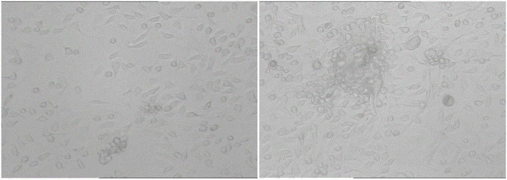
[0043] The result is as figure 1 As shown, as the modeling time prolongs, a certain number of tumor cells that can be detected by the live imager are formed in the abdominal cavity of the mouse.

[0044] 3. Laparotomy imaging

[0045] Intraperitoneal injection of 10*5 cells / only for modeling. After 21 days of modeling, the mice were sacrificed, and the mesentery was imaged.

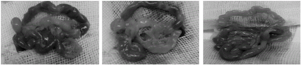
[0046] The result is as figure 2 shown. It can be seen that the mesenteric nodules of the mice are obvious, and obvious ascites is seen. The above results show that: 22 days after the ...

Embodiment 2

[0055] Example 2 Establishment of peritoneal metastasis model by in situ inoculation of cecum wall

[0056] 1. Animal model

[0057] In situ inoculation of cecum wall: 2*10 5 Cells per Matrigel were fixed on the serosa layer of the cecal wall.

[0058] 2. In vivo fluorescence imaging

[0059] Establishment of a peritoneal metastasis model inoculated with the cecal wall and detection of tumor formation and progression using intravital fluorescence imaging.

[0060] The result is as Figure 6 As shown, the results showed that a series of visceral metastases such as liver metastases and lung metastases appeared in mice 17-30 days after injection.

[0061] 3. Laparotomy imaging

[0062] The cecum wall was inoculated with 10*5 cells / only for modeling. After 21 days of modeling, the mice were sacrificed, and the mesentery was imaged.

[0063] The result is as Figure 7 As shown, it can be seen that the peritoneal wall and mesentery metastasis nodules in mice are obvious, and...


PUM


Abstract

The invention relates to a colorectal cancer peritoneal metastasis model and an establishment method thereof. The colorectal cancer peritoneal metastasis model comprises a step (1) of adopting lentivirus transfection to construct HCT116 cells with firefly luciferase genes, so that the HCT116 cells can give out near-infrared light with a wavelength of 500-600 nm under substrate excitation; a step (2) of injecting 10*5 cells into the abdominal cavity of one mouse to perform molding; a step (3) of positioning a tumor by means of D-luciferin in vivo, and observing the carcinogenesis process in real time. The colorectal cancer peritoneal metastasis model has the advantages that a sensitive and accurate in vivo fluorescence imaging system is adopted to evaluate in vivo tumor forming and transfer process in real time, the conventional determination of success in model establishment through mouse dissection is avoided, and the disease pathology progress is observed timely better.

Description

technical field [0001] The invention relates to the field of biotechnology, in particular to a colorectal cancer peritoneal metastasis model and its establishment method. Background technique [0002] Colon cancer is one of the common malignant tumors of the digestive tract. Its incidence rate is second only to lung cancer, gastric cancer, liver cancer, and esophageal cancer in my country. Colon cancer is the third most common tumor in the United States, with an estimated 149,000 new cases and nearly 50,000 deaths from colon cancer in 2008. At present, the incidence of colorectal cancer in my country is still in the process of rising year by year, and the annual new cases increase by about 2%. The standardized incidence rate of colon cancer has risen rapidly. Compared with 1972, the standardized incidence rate in 2000 increased by 134% for males and 135% for females. [0003] Due to the particularity of human beings and ethical considerations, animal models that are closer ...

Claims


Application Information

Patent Timeline
IPC IPC(8): C12N15/867A01K67/027A61K49/00
CPCC12N15/86A01K67/0271A01K2227/105A01K2267/0331A61K49/0045C12N2740/15043
Inventor 蔡国响徐烨蔡三军孟献珂郑洪途李清国
Owner FUDAN UNIV SHANGHAI CANCER CENT
Why Eureka
Try Eureka