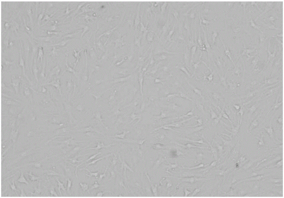
Preparation method of human primary cartilage cells with high yield rate
A high-yield technology for chondrocytes, applied in cell dissociation methods, bone/connective tissue cells, biochemical equipment and methods, etc., can solve the problem of low yield of chondrocytes and achieve the effect of improving the yield
- Summary
- Abstract
- Description
- Claims
- Application Information
AI Technical Summary
Problems solved by technology
Method used
Image
Examples
Embodiment 1
[0023] In this embodiment 1, based on the needs of the implementation process, the instruments and equipment used in the implementation are as follows:
[0024] HH-6 digital display constant temperature water bath (Jintan Fuhua Instrument Co., Ltd.), Varioskan Flash full-wavelength multi-functional microplate reader (Thermo, USA), 3K30 refrigerated high-speed centrifuge (Sigma, Germany), 5450 small High-speed centrifuge (Eppendorf, Germany), Class II Type B2 biological safety cabinet (Esco, Singapore), CO 2 Cell incubator (Thermo, USA), Cellometer Mini automatic cell counter (Nexcelom, USA), DMI8 inverted fluorescence microscope (Leica, Germany).
[0025] The implementation process proceeds in the following steps:
[0026] With the approval of the Ethics Committee and the patient’s informed consent, the knee articular cartilage tissue of the joint replacement patient was taken as the cultured chondrocytes; the obtained cartilage tissue was weighed, put into DPBS buffer (15mL ...
Embodiment 2
[0028] In this embodiment, based on the needs of the implementation process, the instruments and equipment used in the implementation are as follows:
[0029] HH-6 digital display constant temperature water bath (Jintan Fuhua Instrument Co., Ltd.), Varioskan Flash full-wavelength multi-functional microplate reader (Thermo, USA), 3K30 refrigerated high-speed centrifuge (Sigma, Germany), 5450 small High-speed centrifuge (Eppendorf, Germany), Class II Type B2 biological safety cabinet (Esco, Singapore), CO 2 Cell incubator (Thermo, USA), Cellometer Mini automatic cell counter (Nexcelom, USA), DMI8 inverted fluorescence microscope (Leica, Germany).
[0030] The implementation process proceeds in the following steps:
[0031] With the consent of the Ethics Committee and the patient’s informed consent, the cartilage tissue from the knee joint replacement patient was taken as cultured chondrocytes; the obtained cartilage tissue was shaken and washed 3 times in DPBS buffer (15mL cent...
Embodiment 3
[0033] In this embodiment, based on the needs of the implementation process, the instruments and equipment used in the implementation are as follows:
[0034] HH-6 digital display constant temperature water bath (Jintan Fuhua Instrument Co., Ltd.), Varioskan Flash full-wavelength multi-functional microplate reader (Thermo, USA), 3K30 refrigerated high-speed centrifuge (Sigma, Germany), 5450 small High-speed centrifuge (Eppendorf, Germany), Class II Type B2 biological safety cabinet (Esco, Singapore), CO 2 Cell incubator (Thermo, USA), Cellometer Mini automatic cell counter (Nexcelom, USA), DMI8 inverted fluorescence microscope (Leica, Germany).
[0035] The implementation process proceeds in the following steps:
[0036]With the consent of the Ethics Committee and the patient’s informed consent, the cartilage tissue from the knee joint replacement patient was taken as cultured chondrocytes; the obtained cartilage tissue was shaken and washed 3 times in DPBS buffer (15mL centr...
PUM
 Abstract
Description
Claims
Application Information
 - Generate Ideas
- Intellectual Property
- Life Sciences
- Materials
- Tech Scout
- Unparalleled Data Quality
- Higher Quality Content
- 60% Fewer Hallucinations
Browse by: Latest US Patents, China's latest patents, Technical Efficacy Thesaurus, Application Domain, Technology Topic, Popular Technical Reports.
© 2025 PatSnap. All rights reserved.Legal|Privacy policy|Modern Slavery Act Transparency Statement|Sitemap|About US| Contact US: help@patsnap.com

