Reconfigurable X-ray Energy Spectrum Detection Method and Detector Pixel Unit Structure

A detector pixel and unit structure technology, applied in the field of medical X-ray energy spectrum detection, can solve the problems of insufficient energy discrimination of energy spectrum CT detectors, and achieve the effect of improving quality and reducing radiation dose

Inactive Publication Date: 2018-12-07
TIANJIN UNIV
View PDF5 Cites 0 Cited by
  • Summary
  • Abstract
  • Description
  • Claims
  • Application Information

AI Technical Summary

Problems solved by technology

[0004] In order to overcome the deficiencies of the existing technology, solve the problem of insufficient energy discrimination of existing energy spectrum CT detectors, and improve the accuracy of energy spectrum CT imaging, the present invention proposes a high-precision X-ray energy spectrum detector pixel structure and Corresponding spectral layered detection method

Method used


Image

  • Reconfigurable X-ray Energy Spectrum Detection Method and Detector Pixel Unit Structure
  • Reconfigurable X-ray Energy Spectrum Detection Method and Detector Pixel Unit Structure
  • Reconfigurable X-ray Energy Spectrum Detection Method and Detector Pixel Unit Structure

Examples


Embodiment Construction

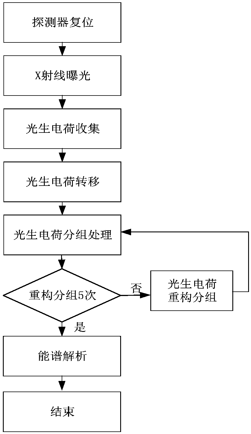
[0024] When the composite X-ray passes through the semiconductor, due to the photoelectric effect, the Compton effect and the electron pair effect, the X-ray (that is, the incident photon) will be absorbed in the semiconductor and generate photogenerated charges, and the X-ray intensity of different energies follows an exponential decay. As a rule, low-energy X-rays will be absorbed first, and high-energy X-rays will be absorbed more slowly. Based on the above X-ray absorption law, the present invention proposes an edge-incidence type X-ray energy spectrum detector pixel structure, and proposes a method for detecting X-ray segmented energy spectrum information using the pixel structure.

[0025] The specific embodiment of the detector pixel structure proposed by the present invention is as follows:

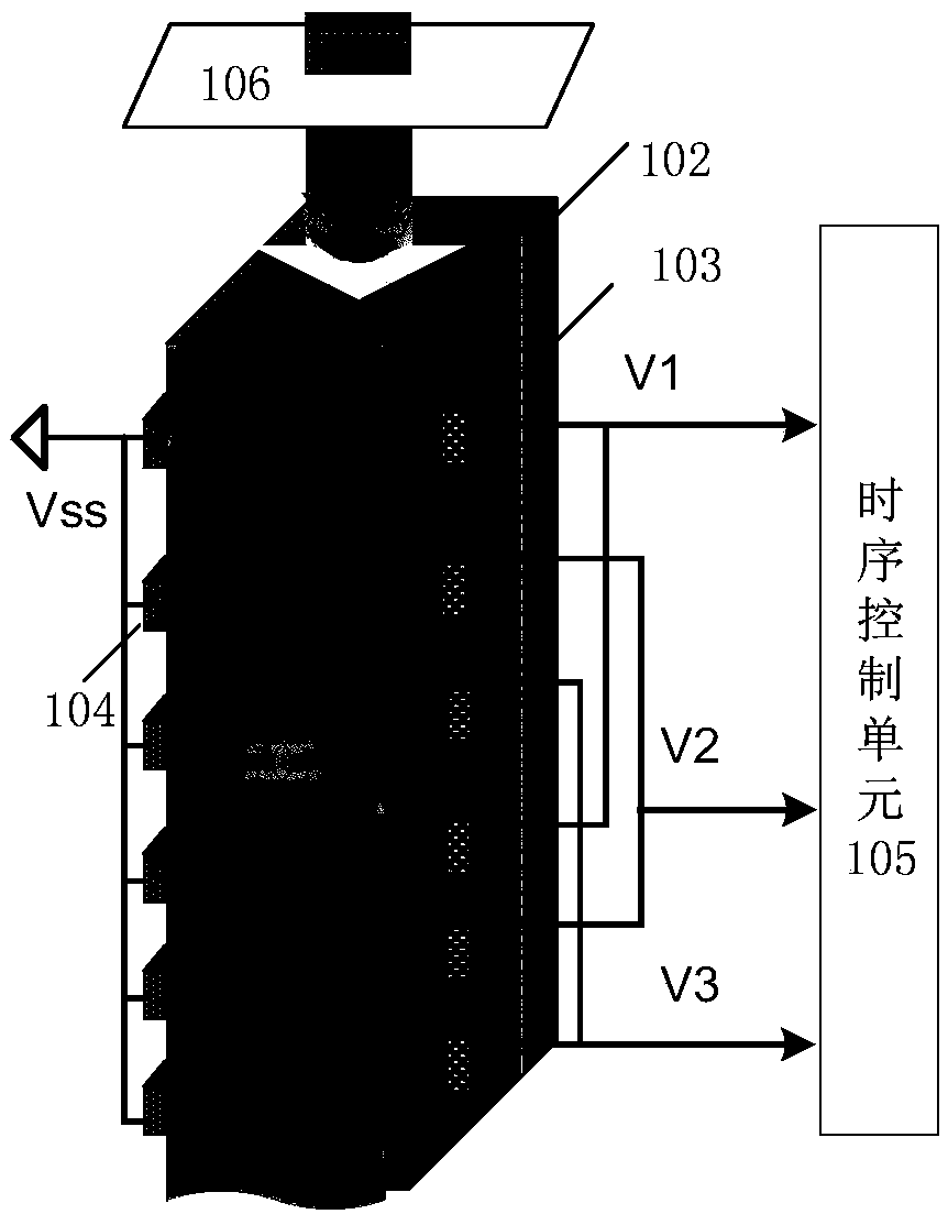
[0026] The basic 3D detector pixel structure is as figure 1 As shown, 101 is the substrate part of the detector. The substrate material can be lightly doped P-type silicon, or ot...


PUM


Abstract

The invention relates to a reconstructible X ray power spectrum detection method and a pixel unit structure of a reconstructible X ray power spectrum detector, belongs to the field of semiconductor photoelectric detectors, and aims at solving the problem that a present power energy CT detector is insufficient in the energy distinguishing degree and improving the precision of power spectrum CT imaging. Attenuation information of X rays of different energy segments can be obtained via once exposure. The pixel unit structure, as a substrate part of a detector body, is used to receive X rays and convert photoelectric signals, and the substrate part is in a size in which all photons of X rays within a to-be-detected energy spectrum range are absorbed two surfaces of the substrate part include oxides of thermal oxide growth, and the substrate part is electrically insulated and isolated from electrodes via the oxides; the electrodes are connected to a time sequence control unit via metal leads, and charge packets are read out sequentially; and a light shield plate which is used to control exposure time of the X rays is provided. The detection method and pixel unit structure are mainly applied to design and manufacture of the semiconductor photoelectric detector.

Description

technical field [0001] The invention relates to the field of semiconductor photodetectors, in particular to the detection of medical X-ray energy spectrum. Background technique [0002] The detectors of traditional medical spiral CT (Computed Tomography, computerized tomography) are detected based on the average attenuation effect of X-rays. The reconstructed image is seriously affected by the beam hardening effect and cannot distinguish similar tissue structures in the human body. At present, with the continuous development of detector technology, spectral CT, which is more sensitive to substances, has increasingly become the focus of research. Spectral CT is based on the difference in the absorption capacity of different photon energies of human tissues, and uses the attenuation characteristics of multiple groups of X-ray energy for image reconstruction, which can provide more image information than conventional CT. The key technology of spectral CT imaging is to use dete...

Claims


Application Information

Patent Timeline
Patent Type & Authority Patents(China)
IPC IPC(8): G01T1/24H01L31/115
CPCG01T1/24H01L31/115
Inventor 史再峰孟庆振杨浩宇曹清洁张嘉平
Owner TIANJIN UNIV
Why Eureka
Try Eureka