
PLGA-S1P nanometer material and preparation method of PLGA-S1P nanometer coating stent

A technology of PLGA-S1P and nanomaterials, which is applied in coating, medical science, surgery, etc., can solve the problems of destroying vascular endothelialization, delaying the natural healing of blood vessels, etc., and achieve reduction of medical expenses, huge economic and social benefits, and improvement prognostic effect

Active Publication Date: 2016-06-29
THE FIRST HOSPITAL OF CHINA MEDICIAL UNIV
View PDF3 Cites 2 Cited by
  • Summary
  • Abstract
  • Description
  • Claims
  • Application Information

AI Technical Summary

Problems solved by technology

However, the drug-eluting stents that have been used clinically can inhibit the proliferation of smooth muscle cells and endothelial cells at the same time, destroying vascular endothelialization and delaying the natural healing of blood vessels.

Method used

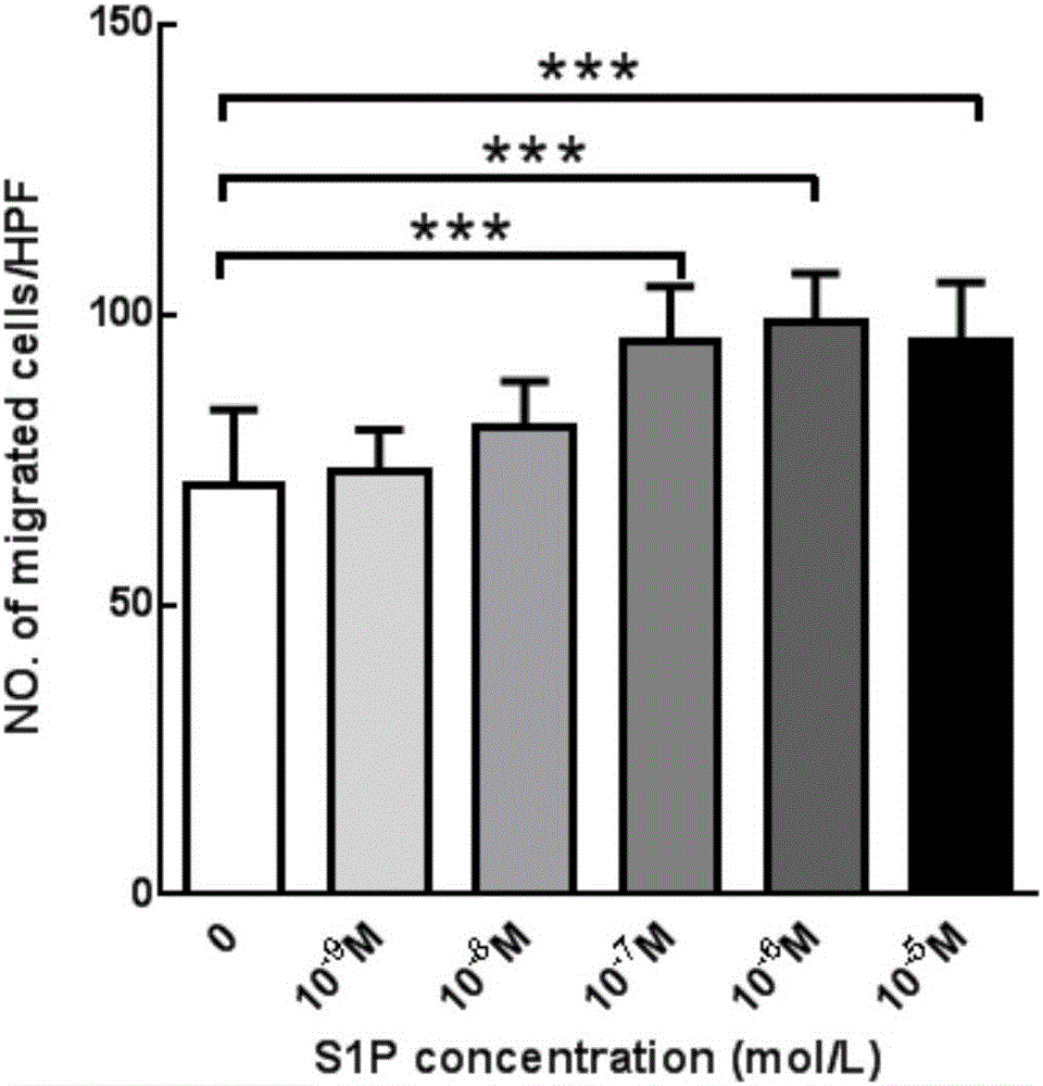
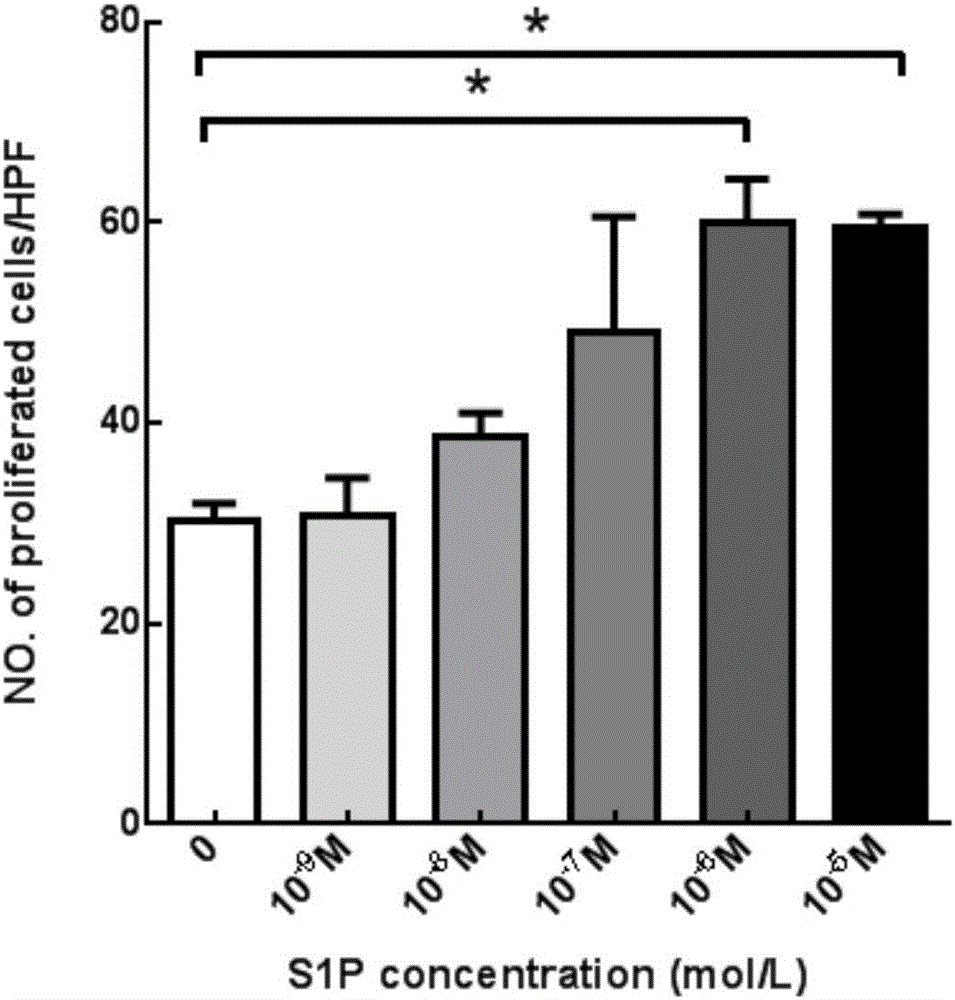
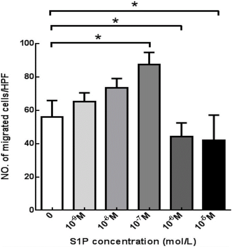

Image

  • PLGA-S1P nanometer material and preparation method of PLGA-S1P nanometer coating stent
  • PLGA-S1P nanometer material and preparation method of PLGA-S1P nanometer coating stent
  • PLGA-S1P nanometer material and preparation method of PLGA-S1P nanometer coating stent

Examples


Embodiment 1

[0028] 1. Preparation of PLGA-S1P nanoparticles:

[0029] 1. Dissolve 100mg PLGA5005 in 4ml dichloromethane organic solvent to prepare PLGA dichloromethane solution.

[0030] 2. Dissolve 1mg S1P in 5ml methanol solution to make S1P solution.

[0031] 3. Under the ultrasonic condition of 180w probe, drop the S1P solution (peristaltic pump 20r / min) into the PLGA dichloromethane solution (ice bath), and ultrasonicate for 3 minutes after dropping to form an emulsion.

[0032] 4. Under 180W probe ultrasound, pour 10ml of 2% PVA (30,000-70,000, degree of alcoholysis 87%-89%, Sigma) solution into the emulsion obtained in step 3 above, and ultrasound for 7 minutes (ice bath).

[0033] 5. Under ultrasound with 180W probe, add the emulsion of step 4 dropwise to 50mL 0.1% PVA solution (peristaltic pump 40r / min), and ultrasound for 6min after dropwise addition.

[0034] 6. Rotary steaming at 30°C for 30 minutes.

[0035] 7. Rotate speed 12000r / min, time 10min, clean with deionized water three times. ...

Embodiment 2

[0041] 1. Preparation of PLGA-S1P nanoparticles:

[0042] 1. Dissolve 10mg PLGA7505 in 4ml tetrachloromethane organic solvent to prepare PLGA tetrachloromethane solution.

[0043] 2. Dissolve 1mg S1P in 5ml acetone solution to make S1P acetone solution.

[0044] 3. Under the ultrasonic condition of 180w probe, add 5mL S1P acetone solution (peristaltic pump 20r / min) dropwise to 4ml PLGA tetrachloromethane solution (ice bath), and ultrasonicate for 5min after dropping to form an emulsion.

[0045] 4. Under 180W probe ultrasound, pour 20ml of 5% PVA (30,000-70,000, degree of alcoholysis 87%-89%, Sigma) solution into the emulsion obtained in step 3 above, and ultrasound for 10min (ice bath).

[0046] 5. Under 180W probe ultrasound, add the emulsion of step 4 dropwise to 60mL of 1% PVA solution (peristaltic pump 100r / min), and ultrasound for 8min after dropwise addition.

[0047] 6. Rotary steaming at 30°C for 30 minutes.

[0048] 7. Rotate speed 10000r / min, time 6min, clean with deionized wat...


PUM

PropertyMeasurementUnit
thicknessaaaaaaaaaa
thicknessaaaaaaaaaa
thicknessaaaaaaaaaa

Abstract

The invention aims at providing a PLGA-S1P nanometer material and a preparation method of a PLGA-S1P nanometer coating stent. The preparation method is characterized by comprising the following steps that S1P is subjected to PLGA coating by a solvent emulsifying method by using an ultrasonic dispersion method, and the ratio of the S1P to PLGA is 1:(10 to 200) (w / w); then, an organic solvent is removed by a method of diffusing an emulsifying agent to water; and finally, freeze-dried powder is prepared by using a freeze-drying concentration method. The PLGA-S1P nanometer particle freeze-dried powder is added into the solvent; after the ultrasonic dissolution, the solution is used as a medicine carrying coating solution; a stainless steel stent is cleanly cleaned; the medicine carrying coating solution is repeatedly sprayed and coated on the surface of the stent by a high-voltage electric spinning method / an ultrasonic spray coating technology; the coating thickness is controlled to be 5 to 20 <mu>m; a de-ionized water / organic solvent volatilization method is used for cleaning; and the PLGA-S1P nanometer coating stent is obtained after the sterilization. The PLGA-S1P nanometer particles are used as the surface coating material of a metal bare stent; and the goal of inhibiting the in-stent restenosis can be achieved. The invention provides a novel method for the interventional therapy for preventing arteriostenosis / occlusion diseases.

Description

Technical field [0001] The invention relates to the field of medical devices, and in particular provides a method for preparing a PLGA-S1P nano material and a PLGA-S1P nano-coated stent. Background technique [0002] Arterial stenosis / occlusion has seriously endangered people's health and has gradually become one of the most important public health problems that humans must face at present and in the future. [0003] Stent implantation has become the first choice for the treatment of peripheral arterial stenosis / occlusive diseases caused by atherosclerosis due to its advantages such as less patient pain, high surgical success rate, fewer complications, quick postoperative recovery, and short hospital stay. Although vascular stent implantation has many advantages as a new technology, the occurrence of in-stent restenosis (ISR) after stent implantation has become a more difficult problem, which has seriously affected the medium and long-term efficacy of interventional therapy. , And...

Claims


Application Information

Patent Timeline
Patent Type & Authority Applications(China)
IPC IPC(8): A61L31/10A61L31/14
CPCA61L31/10A61L31/148A61L2420/02C08L67/04
Inventor 戚勋钟红珊徐克苑永辉
Owner THE FIRST HOSPITAL OF CHINA MEDICIAL UNIV
Learn More