Cancer Markers
- Summary
- Abstract
- Description
- Claims
- Application Information
AI Technical Summary
Benefits of technology
Problems solved by technology
Method used
Image
Examples
example 1
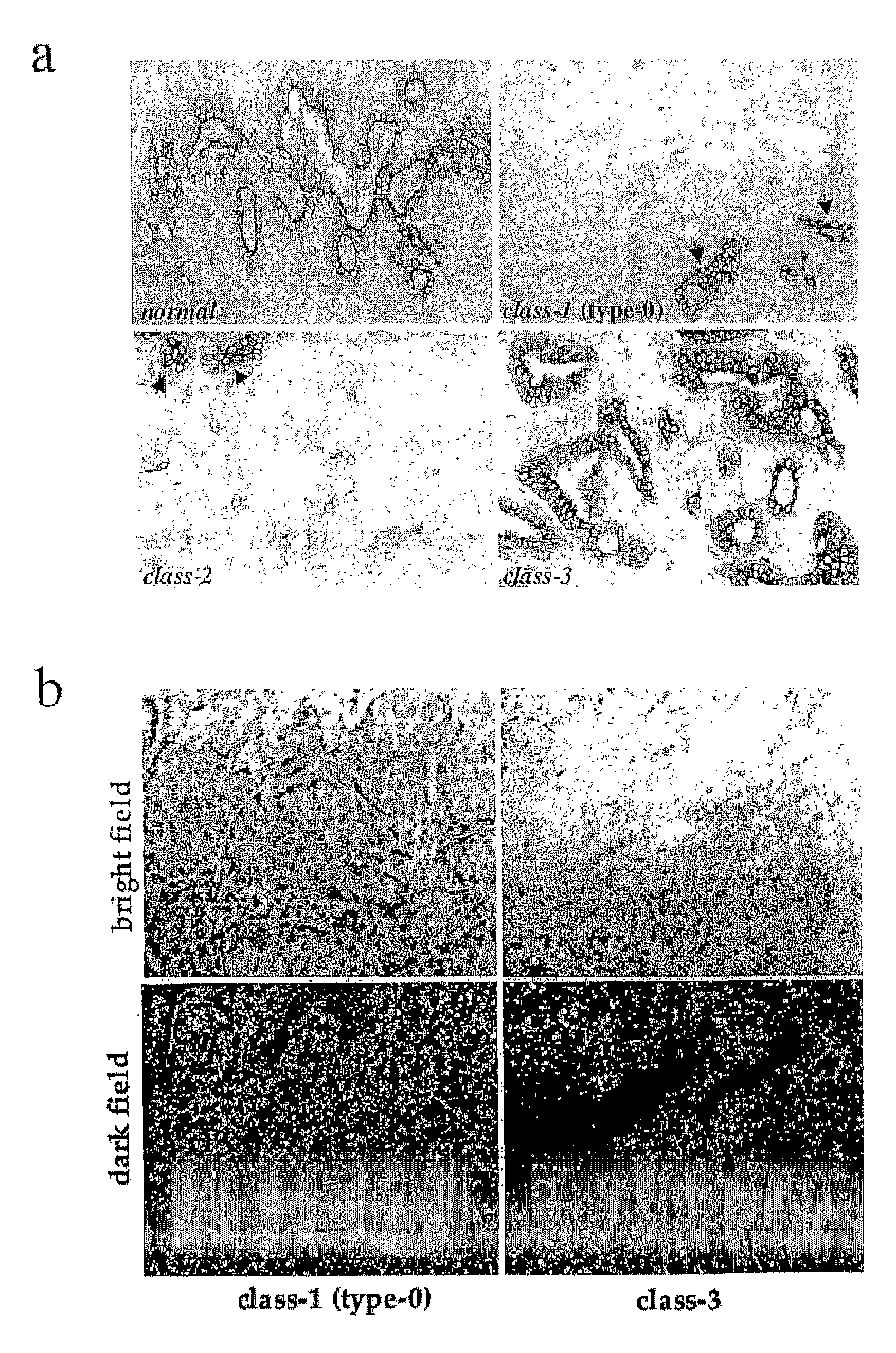
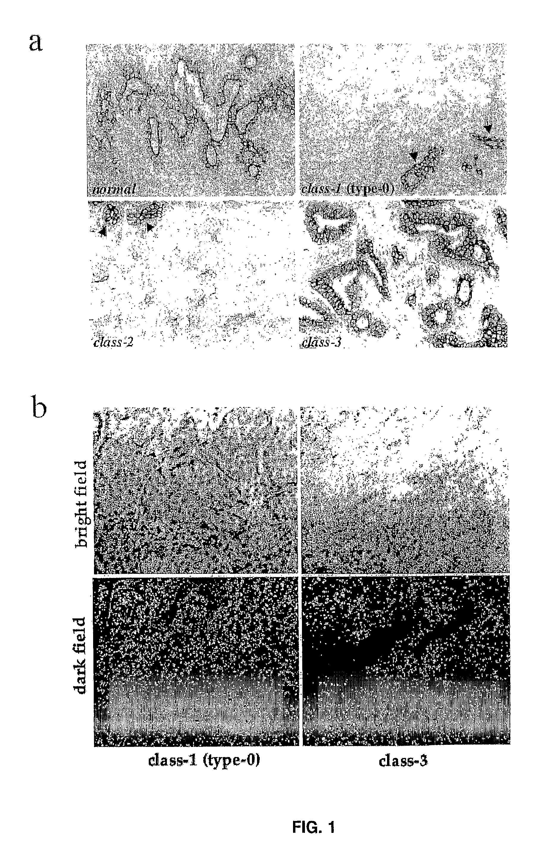
[0409]We characterised by immunohistochemistry 321 consecutive breast cancers. The clinical and pathological features of the breast cancer patients are shown below.
TABLE 1VariableFrequencyPercentAge 0-393912.440-498226.150-59 99229.360-697724.570+247.6HistotypeDuctal24977.6Lobular3811.8Other3410.6Stage (pTclassification)pT120664.2pT29529.6pT3-4206.2Grade17021.8212438.6312739.6Oestrogen receptor7523.4>10%24676.6Progesteronereceptor14846.1>10%17353.9Ki-6714745.8>22%17454.2Lymph nodemetastasisNEG23172.0POS9028.0
[0410]Archival formalin-fixed, paraffin-embedded surgical specimens from the patients were analysed for Numb expression by immunohistochemistry. Tumours were histologically classified according to WHO Histological Classification of Breast Tumours (WHO. Histological typing of breast tumours, 2nd ed., international histological classification of tumours no. 2. Geneva: WHO; 1981) as modified by Rosen and Oberman (Rosen, P. P., Oberman, H. A. Tumours of the mammary gland. Washington...
example 2
[0413]We next analysed the presence of Numb transcripts in human mammary tumours, by in situ hybridisation. Five class-3 tumours, and 14 class-1 (type-0) tumours were analysed. All of the class-3 tumours (and normal glands surrounding the tumours) displayed readily detectable levels of Numb transcripts (FIG. 1b). Interestingly, 12 of 14 class-1 (type-0) tumours displayed levels of Numb mRNA expression comparable to those detected in normal tissues and in class-3 tumours (FIG. 1b).
[0414]In addition, we could not detect any genetic alteration, affecting the Numb locus by both analysis of loss of heterozigosity, and by direct sequencing of Numb cDNAs prepared from several Numb-negative tumours. Thus, genetic alterations at the Numb locus are unlikely to account for lack of Numb expression in the human breast tumours examined.
[0415]Loss of heterozygosity was assessed according to the following method.
[0416]7 polymorphic STS markers (D14S 71, D14S77, D14S268, D14S277, D14S43, D14S70 and ...
example 3
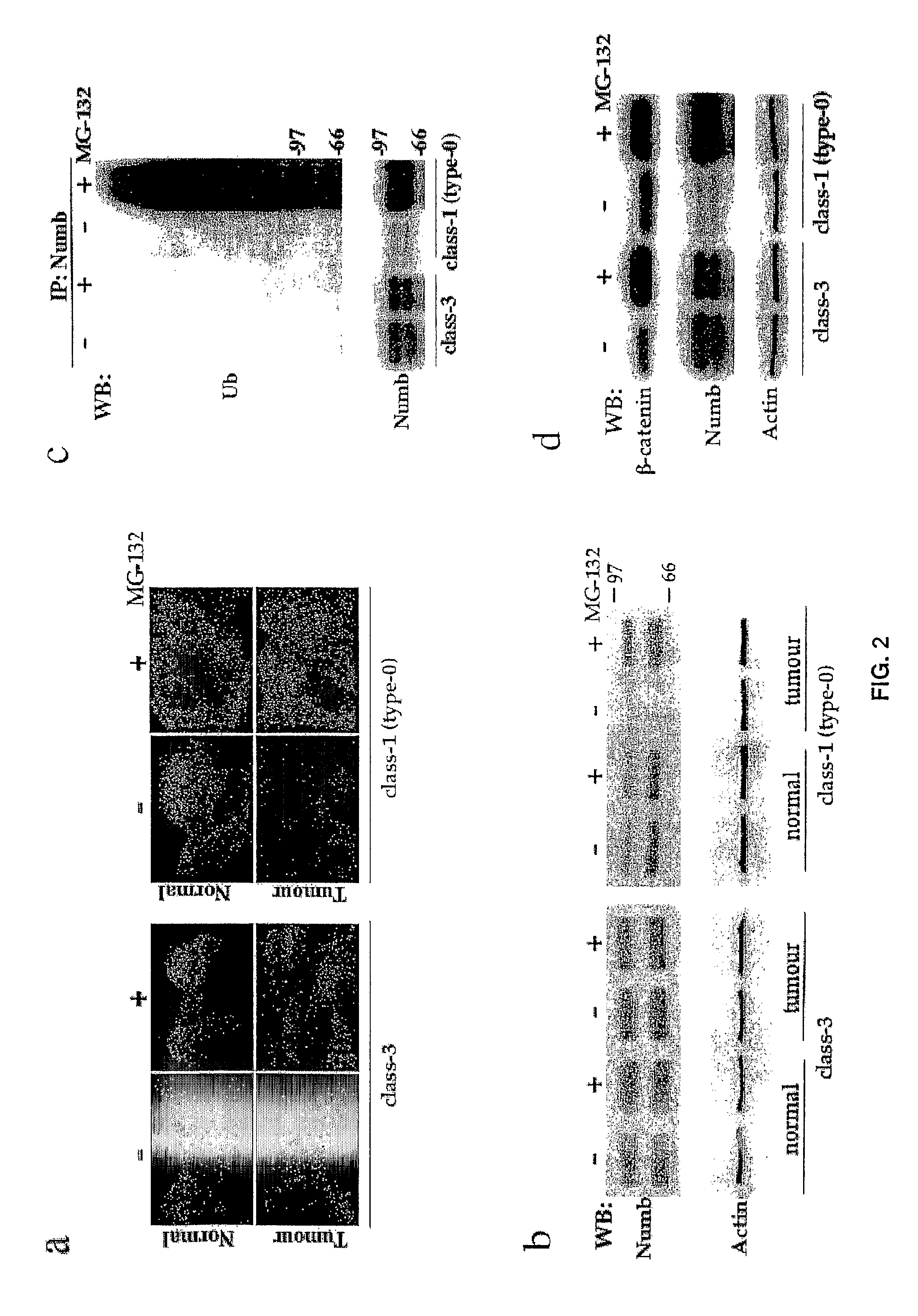
[0419]To gain insight into the molecular mechanisms responsible for loss of Numb expression, we established primary cultures from class-1 (type-0) and class-3 mammary tumours, and from normal breast tissues from the same patients, and analysed them within the first two passages in vitro, as follows.
[0420]Normal and tumour mammary epithelial cells were grown in appropriate selective medium, as described in Methods. Normal cell cultures typically show two major morphological types of cells (top-left): small, smooth-edged, refractile, polygonal cells, which maintain active proliferation and seemingly represent some form of “stem cell population”; and larger, flatter, irregular shaped cells, which do not seem to undergo many cell divisions. The latter might represent cells already programmed to stop multiplying after a few more population doublings. Tumour cells usually display a much higher morphological heterogeneity (top-right), ranging from a typical epithelial appearance to an irre...
PUM
| Property | Measurement | Unit |
|---|---|---|
| Volume | aaaaa | aaaaa |
| Fraction | aaaaa | aaaaa |
| Fraction | aaaaa | aaaaa |
Abstract
Description
Claims
Application Information
 - R&D
- Intellectual Property
- Life Sciences
- Materials
- Tech Scout
- Unparalleled Data Quality
- Higher Quality Content
- 60% Fewer Hallucinations
Browse by: Latest US Patents, China's latest patents, Technical Efficacy Thesaurus, Application Domain, Technology Topic, Popular Technical Reports.
© 2025 PatSnap. All rights reserved.Legal|Privacy policy|Modern Slavery Act Transparency Statement|Sitemap|About US| Contact US: help@patsnap.com


