Examination space for performing computed tomography images

A tomography and examination room technology, applied in the field of examination rooms, can solve the problems of inability to ensure continuous patient presence, insufficiency of patients, and interruption of examinations, and achieve the effects of reducing heartbeat and respiration, improving image quality, and reducing installation costs.

Active Publication Date: 2016-03-02
SIEMENS HEALTHCARE GMBH
View PDF8 Cites 2 Cited by
  • Summary
  • Abstract
  • Description
  • Claims
  • Application Information

AI Technical Summary

Problems solved by technology

[0007] In classic X-ray examinations, mammography or in the case of digital volume tomography (DVT), constant proximity to the patient cannot be ensured, since here at least the individual recordings are initiated by actuating a separate switch outside the examination room. ongoing
[0008] Such a spatial separation of the patient from the medical or operator leads to increased unreliability which the patient mostly always perceives in such examinations
Correspondingly, it can happen that the patient to be examined is not able to follow the breathing instructions given to him sufficiently or even at all (non-cooperation of the patient), which in the worst case leads to an interruption of the examination

Method used


Image

  • Examination space for performing computed tomography images
  • Examination space for performing computed tomography images
  • Examination space for performing computed tomography images

Examples


Embodiment Construction

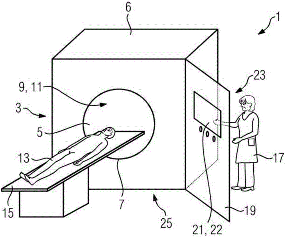
[0049] figure 1 shows an examination room 1 with an imaging device 3 designed as a computed tomography system. The computed tomography system 3 includes an acquisition field 5 with a housing 6 and a gantry 7 . An x-ray radiation source 9 and a data acquisition unit 11 (detector) are arranged in the gantry 7 for the acquisition of x-ray radiation.

[0050] For this purpose, the patient 13 which has been moved into the gantry 7 on the patient table 15 can be positioned in the gantry 7 . The radiation source 9 is rotated around the patient 13 during the examination, emitting a thin X-ray beam while rotating. Each body segment of patient 13 is thus scanned individually.

[0051] If the patient 13 is moved slowly through the computed tomography system 3 , the medical staff 17 , that is to say the operator, acquires a set of sectional images of the desired body region slice by slice. A detailed three-dimensional view of the individual organs can then be reconstructed from the ac...


PUM


Abstract

The invention relates to an examination space (1, 33, 61, 101, 132) for performing computed tomography images, in which an imaging device (3, 31, 63, 103, 131) for generating image recordings of a patient, a radiation protection wall (19, 43, 65, 112, 135) for protecting operator (17, 87) for performing image recording, and an operating unit (21, 45, 67, 113) are arranged 137) in the examination space, wherein the operating unit (21, 45, 67, 113, 137) is set up and adapted to input the scanning settings, output received image data, and process the captured image data.

Description

technical field [0001] The invention relates to an examination room for performing computed tomography recordings. Background technique [0002] Imaging methods such as computed tomography (CT) are used in the medical field to determine high-resolution cross-sectional images of all body regions and organs of a person. In the case of computed tomography (CT), slices of the body or of the body section to be examined are imaged by means of x-ray radiation. In contrast to conventional x-ray recordings, in computed tomography the body is continuously transmitted through an x-ray beam whose source moves around the patient. [0003] In all imaging methods using X-ray radiation, there are conflicting interests that must be weighed against each other in implementation and operation. On the one hand, the medical professional must be protected against scattered radiation from the imaging device, usually by keeping a certain minimum distance from the device or by corresponding shieldi...

Claims


Application Information

Patent Timeline
Patent Type & Authority Applications(China)
IPC IPC(8): A61B90/00A61B6/03
CPCA61B6/032A61B6/107G21F3/00
Inventor B.多尔富斯M.戈特格斯D.勒奇C.蒂尔费尔德
Owner SIEMENS HEALTHCARE GMBH
Why Eureka
Try Eureka