Ultrasound volume data reconstruction method and system based on dynamic assignment

A technology of dynamic assignment and volume data, applied in the medical field, can solve problems such as poor reconstruction effect and complex algorithm

Active Publication Date: 2021-08-17
嘉恒医疗科技(上海)有限公司
View PDF3 Cites 0 Cited by
  • Summary
  • Abstract
  • Description
  • Claims
  • Application Information

AI Technical Summary

Problems solved by technology

However, the current three-dimensional reconstruction method of ultrasound images has the defects of complex algorithm and poor reconstruction effect.

Method used


Image

  • Ultrasound volume data reconstruction method and system based on dynamic assignment
  • Ultrasound volume data reconstruction method and system based on dynamic assignment
  • Ultrasound volume data reconstruction method and system based on dynamic assignment

Examples


Embodiment Construction

[0026] The present invention will be described in further detail below in conjunction with the accompanying drawings and specific embodiments, but not as a limitation of the present invention.

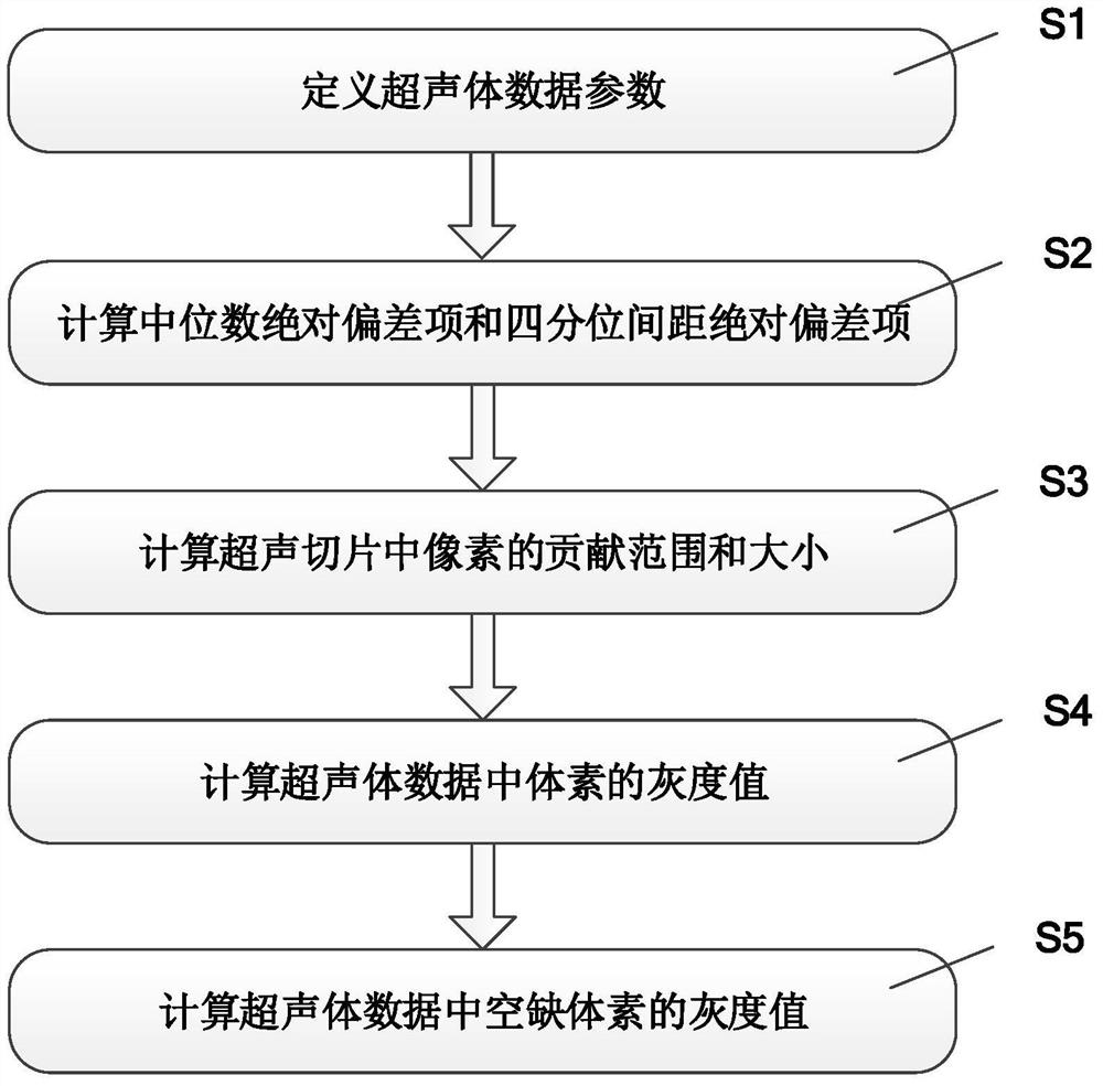
[0027] figure 1 It is a schematic flowchart of a method for reconstructing ultrasound volume data based on dynamic assignment according to an embodiment of the present invention, and the specific steps include:

[0028] Step S1,

[0029] Based on the spatial location of the region of interest, ultrasound volume data parameters are defined. Define the size of the ultrasound volume data as n 1 ×n 2 ×n 3 , the image spacing is [a 1 ,a 2 ,a 3 ], the direction is [x,y,z].

[0030] Among them, the size, direction, and spacing of the ultrasonic volume data can be preset by manually judging the spatial position of the region of interest in the human body.

[0031] Step S2,

[0032] Define the median absolute deviation term and the interquartile range absolute deviation term to calcul...


PUM


Abstract

The invention discloses a method and system for reconstructing ultrasonic volume data based on dynamic assignment. The method includes: defining ultrasonic volume data parameters according to the spatial position of the region of interest; defining the median absolute deviation item and the interquartile range absolute deviation item, and calculating The contribution of the pixel gray level in the ultrasound slice; define the weight of the contribution of the median absolute deviation item and the interquartile range absolute deviation item to the pixel gray level in the ultrasound slice, and calculate the contribution range and size of the pixel in the ultrasound slice; according to the used The pixel contribution range and size of all ultrasound slices reconstructed from the ultrasound volume data, and the gray value of the voxels in the ultrasound volume data are calculated; the gray value of the vacant voxels in the ultrasound volume data is calculated by an interpolation method. The invention can quickly and accurately reconstruct ultrasonic volume data, and provide theoretical guidance for diagnosis and treatment of clinical lesions.

Description

technical field [0001] The invention relates to the field of medical technology, in particular to a method and system for reconstructing ultrasonic volume data based on dynamic assignment. Background technique [0002] Ultrasound imaging is a method and technology that uses ultrasonic waves to irradiate the human body, and obtains visible images of the nature and structure of human tissue by receiving and processing echoes that carry characteristic information of human tissue. Together with CT and MRI, it is called the three major imaging studies of modern medicine. . Compared with CT and MRI, ultrasound imaging has the advantages of short acquisition time, no need for intravenous contrast agent, no ionizing radiation, economical and convenient, real-time imaging, etc., and has become an essential clinical diagnostic and therapeutic equipment. However, when doctors want to understand the structure of organs more accurately, traditional 2D imaging cannot meet the requirement...

Claims


Application Information

Patent Timeline
Patent Type & Authority Patents(China)
IPC IPC(8): G06T17/00A61B8/00
Inventor 郑重吴文波杨文晖赖暖翔
Owner 嘉恒医疗科技(上海)有限公司
Why Eureka
Try Eureka