Eureka AIR delivers breakthrough ideas for toughest innovation challenges, trusted by R&D personnel around the world.

Classifier for micro-angioma of diabetes lesion based on colored image

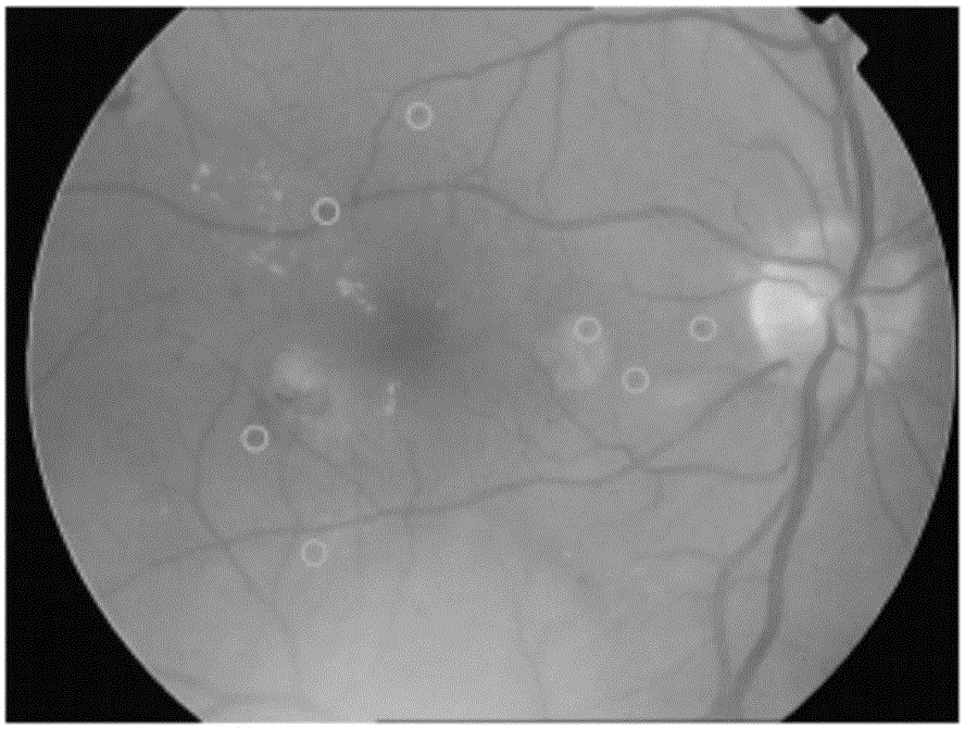
A color image and microangioma technology, which is applied in the field of medical image processing, can solve the problems of misjudgment of small blood vessels as microangioma, discontinuous gray distribution, and influence, and achieve the effect of strong practicability, simple operation, and convenient use

Inactive Publication Date: 2015-11-18
XI AN JIAOTONG UNIV
View PDF5 Cites 20 Cited by
  • Summary
  • Abstract
  • Description
  • Claims
  • Application Information

AI Technical Summary

Problems solved by technology

However, since microvascular tumors are not clear in color retinal images, the accuracy of microvascular tumor detection is affected by background noise, and during filtering operations, microvascular tumors are often surrounded by small blood vessels, which may be "merged" into small blood vessels. Blood vessel
In addition, the gray distribution along the direction of blood vessels is often discontinuous, so small blood vessels are also easily misjudged as microvascular tumors

Method used


Image

  • Classifier for micro-angioma of diabetes lesion based on colored image
  • Classifier for micro-angioma of diabetes lesion based on colored image
  • Classifier for micro-angioma of diabetes lesion based on colored image

Examples


Embodiment Construction

[0033] The present invention is described in further detail below in conjunction with accompanying drawing:

[0034] refer to figure 1 The apparatus for detecting microvascular tumors of diabetic lesion variants based on color images according to the present invention includes an image preprocessing subsystem for denoising the retinal images of candidates, and an image preprocessing subsystem for locating microvascular tumors in the retinal images of candidates. Multi-size, multi-direction and matched filter, region growing subsystem for restoring the size and shape of the microvascular tumor in the retinal image of the candidate, feature extraction for extracting 37-dimensional features of the microvascular tumor in the retinal image of the candidate system, and a support vector machine-based classification subsystem for classifying candidates;

[0035] The output end of the image processing subsystem is sequentially connected with the input end of the classification subsyst...


PUM


Abstract

The invention discloses a classifier for micro-angioma of diabetes lesion based on a colored image. The classifier comprises an image pre-processing subsystem, a multi-size multi-directional matching filter, a regional growth subsystem, a characteristic quantity extraction system and a classification subsystem, wherein the image pre-processing subsystem reduces noise of a retina image of a candidate, the filter positions the micro-angioma in the retina image, the regional growth subsystem restores the size and shape of the micro-angioma in the retina image, the characteristic quantity extraction system extracts 37-dimensional characteristic of the micro-angioma in the retina image, and the classification subsystem classifies the candidate based on a support vector machine; and the output end of the image pre-processing subsystem is connected with the input end of the classification subsystem via the multi-size multi-directional matching filter, the regional growth subsystem and the characteristic quantity extraction system. The classifier can effectively identify the micro-angioma in the retina image.

Description

technical field [0001] The invention belongs to the field of medical image processing, and relates to a color image-based classification device for microvascular tumors of diabetic lesions. Background technique [0002] According to a study by the American Diabetes Association (ADA), from 2005 to 2008, diabetic retinopathy (DR) affected more than 4.4 million Americans over the age of 37, of which nearly 700,000 patients had diabetic retinopathy leading to Possibility of severe vision loss. Early detection and treatment of DR can reduce the risk of severe vision loss by more than 90%. Therefore, a high-efficiency and cost-effective DR screening system has begun to receive widespread attention. The methods used to check DR mainly include ophthalmoscopy, color fundus photography, fundus fluorescein angiography (FFA), ultrasound, and electroretinogram. Among them, color fundus photography has the characteristics of simple, fast and convenient automatic image analysis and proce...

Claims


Application Information

Patent Timeline
IPC IPC(8): G06T7/00G06K9/62
CPCG06T7/0012G06T2207/30041G06T2207/10024G06F18/2411
Inventor 辛景民王庆洁武佳懿郑南宁
Owner XI AN JIAOTONG UNIV
Why Eureka
Eureka Blog
Learn More