
Method and system for diagnosing uterine contraction levels using image analysis

A uterine contraction and uterine technology, applied in image analysis, 2D image generation, diagnosis, etc., can solve problems such as inability to distinguish from contractions, movement of boundaries, and noise.

Inactive Publication Date: 2017-05-31
FERRING BV
View PDF7 Cites 0 Cited by
  • Summary
  • Abstract
  • Description
  • Claims
  • Application Information

AI Technical Summary

Problems solved by technology

A disadvantage of the M-mode approach in detecting uterine contractions is that it does not provide a means of segmenting the entire uterus, only the upper and lower borders that exist within user-specified intersections
The absence of this provision becomes pronounced in the case of exacerbated bowel or respiratory movements that may change the position of the gap in the previously marked uterine border
Additionally, the uterus may move back and forth relative to the intersection that has been set
This shift may appear to be uncorrelated, but it may result in partial graphs that are indistinguishable from shrinkage
Further problems manifest as noise or false edges
Because the method does not take into account the shape of the entire uterus, only boundaries across intersections are traceable and this can make measurements difficult
Even in ideal situations where boundaries are clear and easy to track, accurate measurements can still be difficult
Similarly, it is equally difficult to interpret images where, although contractility is present, the movement of the border remains static and only structural changes in the endometrium are affected

Method used


Image

  • Method and system for diagnosing uterine contraction levels using image analysis
  • Method and system for diagnosing uterine contraction levels using image analysis
  • Method and system for diagnosing uterine contraction levels using image analysis

Examples


Embodiment

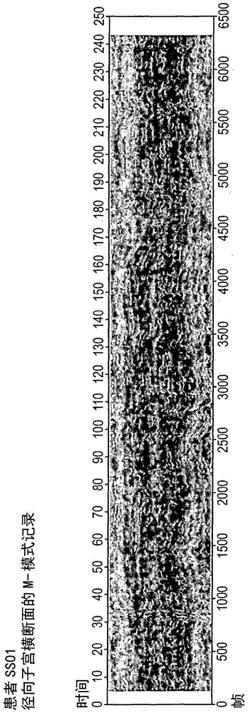
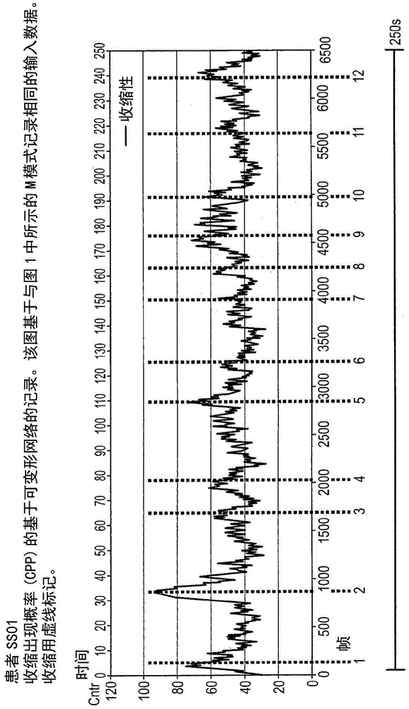
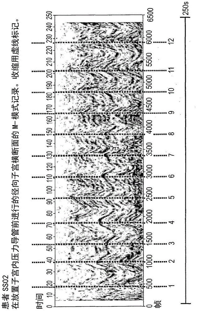
[0082] In order to validate the computer-based deformable model network application method, a clinical study is presented to provide cross-validation of the method against intrauterine pressure measurements. The study involved patients undergoing controlled ovarian stimulation and volunteering for mock embryo transfer (mock ET) and intrauterine pressure assessment. All volunteers underwent controlled ovarian stimulation and had mock ET. Although initially, stimulation cycles in the included patients were designed as treatment cycles, in all cases further treatment was not possible due to exacerbated ovarian response or fertilization failure. After agreeing to the procedure, patients received standard progesterone support (200 mg tid of micronized progesterone, transvaginally). Mock ET and ultrasound scans were performed 2 days after oocyte collection or 2 days + 36 hours after hCG administration in persons who had not initiated oocyte collection. An evaluation in two menstru...


PUM


Abstract

A method for analyzing uterine contractions to support embryo transfer techniques by analyzing uterine images using a deformable model network. The method is also used to diagnose premature uterine contraction activity in mammals. When used with an oxytocin antagonist, the method can be used to control contractile activity during embryo transfer or preterm birth.

Description

Background technique [0001] Detection of uterine contraction activity is necessary in a variety of medical procedures, including embryo transfer during in vitro fertilization, and detection and prevention of preterm birth during pregnancy. [0002] Increased uterine contractility in women undergoing embryo transfer ("ET") may affect ET success rates. Successful implantation rates were up to 3 times higher in ET recipients with a "silent" uterus compared to patients with elevated uterine contractility. It is currently hypothesized that administration of oxytocin antagonists may reduce uterine contractions and may increase pregnancy rates. In transplantation studies conducted in mice, it was confirmed that oxytocin antagonists reversed the negative effects of oxytocin. [0003] In humans, ethical issues preclude the use of invasive techniques for the assessment of uterine contractions, such as intrauterine pressure measurement in patients about to undergo embryo transfer proce...

Claims


Application Information

Patent Timeline
Patent Type & Authority Patents(China)
IPC IPC(8): A61B8/08A61B5/00G06T7/00
CPCA61B5/4356A61B8/08A61B8/485A61B8/5223A61B2576/00G06T2207/10132G06T2207/20116G06T2207/30004G06T7/12G06T7/149A61P15/04G16H50/30A61B17/435A61B2576/02G06T11/206G06T2207/30044G06T2210/41
Inventor 彼得·彼任斯基瓦尔德马·库琴斯基
Owner FERRING BV
Learn More