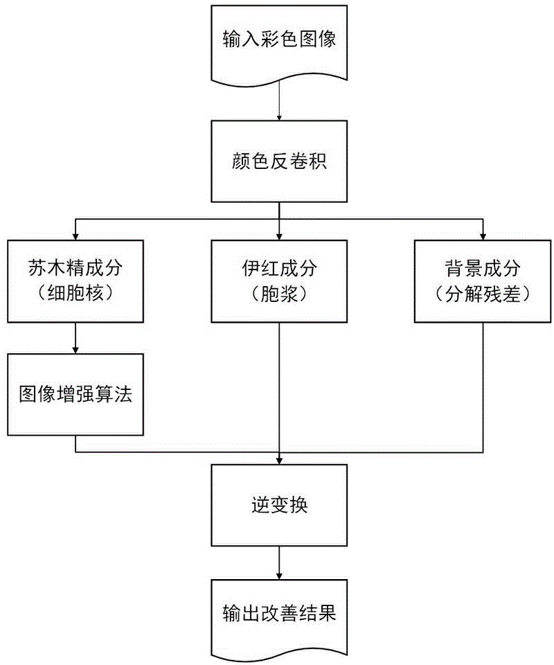
Method for improving pathology image visual effect based on main component analyzing
A principal component analysis, pathological image technology, applied in image enhancement, image data processing, instrumentation, etc., can solve problems such as diagnostic influence, difference, contrast reduction, etc.
- Summary
- Abstract
- Description
- Claims
- Application Information
AI Technical Summary
Problems solved by technology
Method used
Image

Examples
Embodiment 1
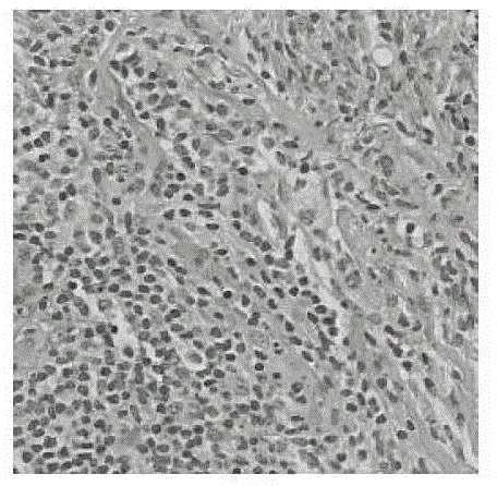
[0059] Such as image 3 as shown, image 3 For the original image of the pathological section in this embodiment, the original image ( image 3 ) image into a matrix form, where i is the abscissa of the image, j is the ordinate of the image, c takes R, G or B to represent the three channels of the color image, and is represented by a matrix D.
[0060] D = ( d ij ) 3 × MN = g 1,1 , R , g 1,2 , R , . . ...
PUM
 Abstract
Description
Claims
Application Information
 - R&D
- Intellectual Property
- Life Sciences
- Materials
- Tech Scout
- Unparalleled Data Quality
- Higher Quality Content
- 60% Fewer Hallucinations
Browse by: Latest US Patents, China's latest patents, Technical Efficacy Thesaurus, Application Domain, Technology Topic, Popular Technical Reports.
© 2025 PatSnap. All rights reserved.Legal|Privacy policy|Modern Slavery Act Transparency Statement|Sitemap|About US| Contact US: help@patsnap.com