
Immobilization device and immobilization method for pathological tissues

A fixation device and pathological technology, which is applied in the field of medical devices, can solve the problems of cross-contamination of pathological tissues, etc., and achieve the effects of avoiding cross-contamination, good fixation effect and consistent effect

Inactive Publication Date: 2015-04-08
GENERAL HOSPITAL OF PLA
View PDF22 Cites 2 Cited by
  • Summary
  • Abstract
  • Description
  • Claims
  • Application Information

AI Technical Summary

Problems solved by technology

However, the soft bag cannot separate each pathological tissue well, so there is a problem of cross-contamination between pathological tissues

Method used


Image

  • Immobilization device and immobilization method for pathological tissues
  • Immobilization device and immobilization method for pathological tissues
  • Immobilization device and immobilization method for pathological tissues

Examples


Embodiment Construction

[0023] The present invention will be further described below in conjunction with accompanying drawing.

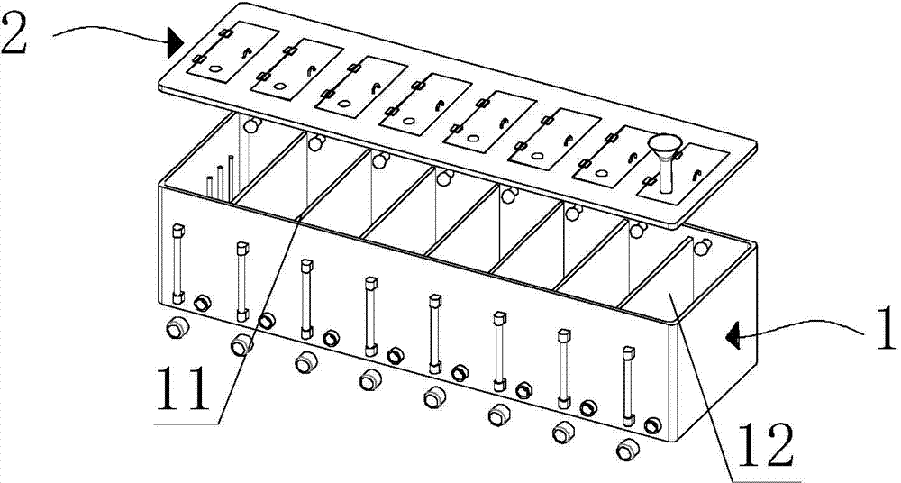
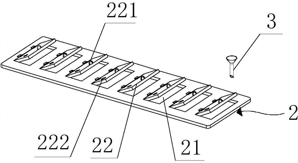
[0024] see figure 1 , which is a schematic diagram of the overall exploded structure of the fixation device for pathological tissue of the present invention. As shown in the figure, the fixing device includes a box body 1 . The box body 1 is used to contain fixative solution and pathological tissue.

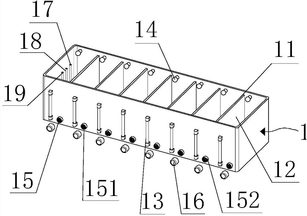
[0025] In the embodiment of the present application, partition members 11 are arranged at intervals in the box body 1 . The partition member 11 divides the box body 1 into a plurality of chambers 12, and each chamber 12 can be filled with fixative solution and pathological tissue. In this way, due to the setting of the partition member 11, the box body 1 is divided into a plurality of chambers 12 independent of each other, so that the purpose of simultaneously fixing multiple pathological tissues can be achieved, and it is also avoided to be located in adjacent chambers. T...


PUM


Abstract

The invention relates to an immobilization device and an immobilization method for pathological tissues. The immobilization device comprises a box body and a box cover assembly, wherein the box body is used for containing a immobilization solution and the pathological tissues; partition components for partitioning the box body into a plurality of chambers are arranged in the middle of the box body at intervals; and the box cover assembly is arranged at an upper end opening of the box body, and is used for sealing the box body. The immobilization device has the advantage of good immobilization effect on the pathological tissues; the cross contamination among the pathological tissues is avoided; and the concentration, the temperature and the pH value of the solution in the immobilization device can be detected in real time.

Description

technical field [0001] The invention relates to the technical field of medical instruments, in particular to a fixing device for pathological tissues and a fixing method thereof. Background technique [0002] In the medical field, in order to study the lesions and the extent of lesions in human organs (such as the stomach, lungs, or breasts, etc.), it is necessary to remove certain sized diseased tissues from the organs that have already undergone lesions. The excised diseased tissue is processed by various chemicals or embedding methods, so that the excised diseased tissue is fixed and hardened. The lesion tissue is cut into thin slices on a microtome, and the thin slices are adhered to a glass slide to achieve position fixation of the thin slices. At the same time, the thin slices are dyed to facilitate the observation of the morphology of pathological tissues under a microscope, so as to make accurate pathological judgments. Therefore, effective fixation of pathological...

Claims


Application Information

Patent Timeline
Patent Type & Authority Applications(China)
IPC IPC(8): G01N1/30
Inventor 袁静王湛博石燕刘洪一石怀银宋欣
Owner GENERAL HOSPITAL OF PLA
Learn More