A method for in situ culture of amniocytes and karyotype processing and analysis on glass slides
A technology of amniotic fluid cells and in situ culture, which is applied to animal cells, vertebrate cells, artificial cell constructs, etc., can solve the problems of high analysis cost and difficult popularization, and achieve high-quality cleavage phase, stable production quality, and high success rate high effect
- Summary
- Abstract
- Description
- Claims
- Application Information
AI Technical Summary
Problems solved by technology
Method used
Image

Examples
Embodiment 1
[0030] Example 1: Synchronous culture of amniocytes on domestic slides and imported slides
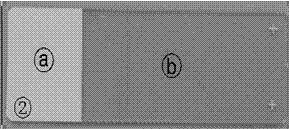
[0031] (1) Inoculation: Select 10 amniotic fluid samples, each sample takes 18-22ml. Aseptically divide into 2 conical centrifuge tubes, 9-11ml / tube, balance and centrifuge for 10min at 1200rpm, keep 0.5ml of cell suspension (amniotic fluid cell pellet + a little amniotic fluid) in each tube, add 1.5ml of amniotic fluid culture medium to each tube , respectively tiled on the cell-adherent surface of the domestic glass slide and the imported original glass slide culture flask ( figure 2 b) at 37°C, 5% CO 2 Incubate for 2 days under conditions with minimal mechanical disturbance, and after 2 days at the mark on the slide ( figure 2 a) Add another 3ml of amniotic fluid medium, mix the newly added medium with the old medium, and place it again at 37°C, 5% CO 2 conditions for 5 days.
[0032] (2) Change the medium: After the amniotic fluid cells have been cultured for 7-8 days, place ...
Embodiment 2
[0047] Example 2: Effects of different hypotonic fluids on cleavage phase during the harvesting of amniotic fluid cultured on domestic slides
[0048] 1) Inoculation: Take 24 amniotic fluid samples, 5ml for each sample, mix them evenly (to eliminate the differences among the samples), divide into 12 bottles, 2 bottles for each condition. 10ml / tube, balance and centrifuge for 10min at 1200rpm, take 0.5ml of cell suspension (amniotic fluid cell pellet + a little amniotic fluid) from each tube, add 1.5ml of amniotic fluid culture medium to each tube, and spread them on domestic glass slides and imported original loading The cell-attached side of the slide culture flask ( figure 2 b) at 37°C, 5% CO 2 Incubate for 2 days under the conditions of minimal mechanical disturbance. After 2 days, mark the slide ( figure 2 a) Add another 3ml of amniotic fluid medium, mix the newly added medium with the old medium, and place it again at 37°C, 5% CO 2 conditions for 5 days.
[0049] 2)...
Embodiment 3
[0055] Example 3: Effects of Different Pre-fixed Solutions on Split Phases During Amniotic Fluid Harvesting in Domestic Slide Culture
[0056] 1) Inoculation: Take 20 amniotic fluid samples, 5ml for each sample, mix them evenly (to eliminate the differences among the samples), divide into 10 bottles, 2 bottles for each condition. 10ml / tube, balance and centrifuge for 10min at 1200rpm, take 0.5ml of cell suspension (amniotic fluid cell pellet + a little amniotic fluid) from each tube, add 1.5ml of amniotic fluid culture medium to each tube, and spread them on domestic glass slides and imported original loading The cell-attached side of the slide culture flask ( figure 2 b) at 37°C, 5% CO 2 Incubate for 2 days under conditions with minimal mechanical disturbance, and after 2 days at the mark on the slide ( figure 2 a) Add another 3ml of amniotic fluid medium, mix the newly added medium with the old medium, and place it again at 37°C, 5% CO 2 conditions for 5 days.
[0057]...
PUM
 Abstract
Description
Claims
Application Information
 - R&D
- Intellectual Property
- Life Sciences
- Materials
- Tech Scout
- Unparalleled Data Quality
- Higher Quality Content
- 60% Fewer Hallucinations
Browse by: Latest US Patents, China's latest patents, Technical Efficacy Thesaurus, Application Domain, Technology Topic, Popular Technical Reports.
© 2025 PatSnap. All rights reserved.Legal|Privacy policy|Modern Slavery Act Transparency Statement|Sitemap|About US| Contact US: help@patsnap.com