Biomedical image feature extraction method

An image feature extraction and biomedical technology, applied in the field of biomedical image feature extraction, can solve the problems of low image classification and recognition accuracy, ignoring image information, etc.

Inactive Publication Date: 2015-01-14
SUN YAT SEN UNIV
View PDF3 Cites 6 Cited by
  • Summary
  • Abstract
  • Description
  • Claims
  • Application Information

AI Technical Summary

Problems solved by technology

[0005] After analyzing the existing technical literature, scholars such as R.Barry and L.Minsuk wrote in "R.Barry, L.Minsuk, Exploring text and image features to classify images in bioscience literature, Proceedings of the Workshop on Linking Natural Language Processing and Biology, 2003, 73-80", proposed a feature extraction method based on the combination of text and vision. This method mainly extracts visual features (features based on histograms, 8-connected region features) and image captions based on biomedical images. Text features, and realize the classification of biomedical images, but these features have low classification and recognition accuracy for images, and ignore the information of text in images

Method used


Image

  • Biomedical image feature extraction method
  • Biomedical image feature extraction method
  • Biomedical image feature extraction method

Examples


Embodiment Construction

[0051] The following will clearly and completely describe the technical solutions in the embodiments of the present invention with reference to the accompanying drawings in the embodiments of the present invention. Obviously, the described embodiments are only some, not all, embodiments of the present invention. Based on the embodiments of the present invention, all other embodiments obtained by persons of ordinary skill in the art without making creative efforts belong to the protection scope of the present invention.

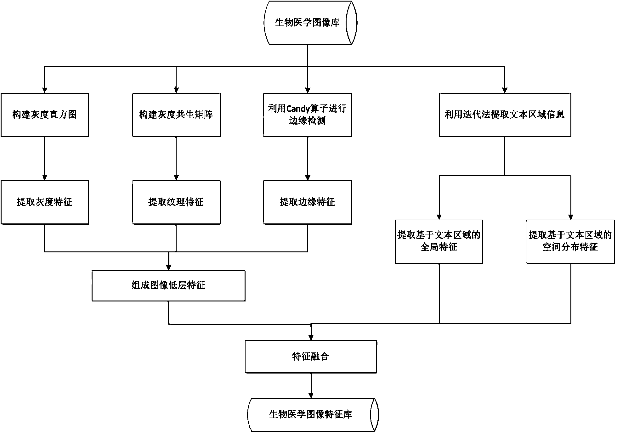
[0052] refer to figure 1 , the biomedical image feature extraction method of the technical solution includes the following steps:

[0053] Step 1: Extract low-level visual features of biomedical image I;

[0054] (1) Use the grayscale histogram to calculate the statistics of the image to obtain the grayscale feature GH of the image;

[0055] (2) Use the gray level co-occurrence matrix to extract the texture feature TH of the image;

[0056] (3) extracting the...


PUM


Abstract

The invention discloses a biomedical image feature extraction method. The method comprises the first step of extracting lower-layer visual features (grey features, textural features and edge features) of a biomedical image, the second step of extracting a text region from the biomedical image to obtain pixel information and position information of the text region, the third step of extracting global features from the text region of the biomedical image, the fourth step of dividing the image into sub-regions through a sub-region division method, analyzing text region distribution of all the sub-regions and extracting spatial distribution features of the image, and the fifth step of conducting feature fusion on the three kinds of image features to obtain final image visual features. By means of the method, the essential properties of the biomedical image can be described comprehensively by analyzing lower-layer visual information and text region information of the image, so the classification accuracy of the biomedical image is effectively improved, and the effect is better when the method is used for identifying flow charts and graphs.

Description

technical field [0001] The invention relates to the field of digital image processing, in particular to a biomedical image feature extraction method. Background technique [0002] With the development of computer multimedia technology and machine learning technology, the application of image database is becoming more and more extensive. How to effectively establish, manage and make full use of image information database resources to realize intelligent and efficient recognition and retrieval of images has always been a hot issue concerned by scientific researchers at home and abroad. In scientific research, national defense, industrial production, aerospace, It has a wide range of applications in biomedicine, traffic monitoring and other fields. Faced with the characteristics of large amount of biomedical image data, multi-modality, complex imaging, and strong pertinence, feature extraction technology is constantly facing new opportunities and challenges [0003] Image fea...

Claims


Application Information

Patent Timeline
Patent Type & Authority Applications(China)
IPC IPC(8): G06T7/00
CPCG06V10/50G06V10/44G06T7/00
Inventor 罗笑南陈湘萍邓伟财王众
Owner SUN YAT SEN UNIV
Why Eureka
Try Eureka