Method for accurately distinguishing cell cycle

A cell cycle, cell technology, applied in the biological field

Inactive Publication Date: 2014-12-17
SHANGHAI INST OF BIOLOGICAL SCI CHINESE ACAD OF SCI
View PDF3 Cites 3 Cited by
  • Summary
  • Abstract
  • Description
  • Claims
  • Application Information

AI Technical Summary

Problems solved by technology

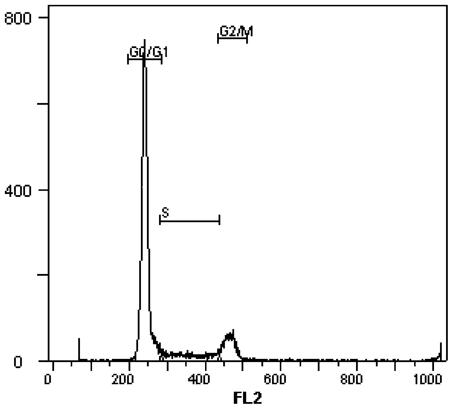
[0003] In summary, there is currently a lack of technology in this field that can distinguish the cell cycle more accurately and in detail, and realize the separation of the G0 phase from the G1 phase, and the separation of the G2 and M phases.

Method used


Image

  • Method for accurately distinguishing cell cycle
  • Method for accurately distinguishing cell cycle
  • Method for accurately distinguishing cell cycle

Examples


Embodiment 1

[0098] Example 1. Accurate cell cycle differentiation of suspension cells

[0099] Taking the analysis of human acute lymphoblastic leukemia cells (Jurkat T) as an example, the method of using the kit of the flow cytometer of the present invention that can accurately locate the cell cycle comprises the following steps:

[0100] 1) Fixed: take about 1×10 7 For each cell, add freshly prepared fixative with a final concentration of 0.5% (w / v), and incubate at 37°C for 15 minutes in a carbon dioxide incubator;

[0101] 2) Washing: centrifuge the cells obtained in step 1) at a speed of 400 g for 5 minutes to remove the supernatant, resuspend the cells in 1 ml of cold phosphate buffer, and mix them into single cells;

[0102] 3) Permeabilization: Centrifuge 400 g of the cell suspension obtained in step (2) to remove the supernatant, add 1 ml of cold anhydrous methanol to the cell pellet; mix well while adding, overnight at -20°C;

[0103] 4) According to the method described in st...

Embodiment 2

[0109] Example 2, Accurate cell cycle differentiation of adherent cells

[0110] Taking the analysis of human liver cancer cell line (SMMG-7721) as an example, the method of using the kit of the flow cytometer of the present invention that can accurately locate the cell cycle comprises the following steps:

[0111] 1) Fixation: cells were digested with trypsin at 37°C for 5 minutes into a single suspension cell, about 1×10 7 For each cell, add freshly prepared fixative with a final concentration of 0.5% (w / v), and incubate at 37°C for 15 minutes in a carbon dioxide incubator;

[0112] 2) Washing: centrifuge the cells obtained in step 1) at a speed of 400g for 5 minutes to remove the supernatant, resuspend the cells in cold phosphate buffer, and mix them into single cells;

[0113] 3) Permeabilization: Centrifuge 400 g of the cell suspension obtained in step (2) to remove the supernatant, add 1 ml of cold anhydrous methanol to the cell pellet; mix well while adding, overnight ...


PUM


Abstract

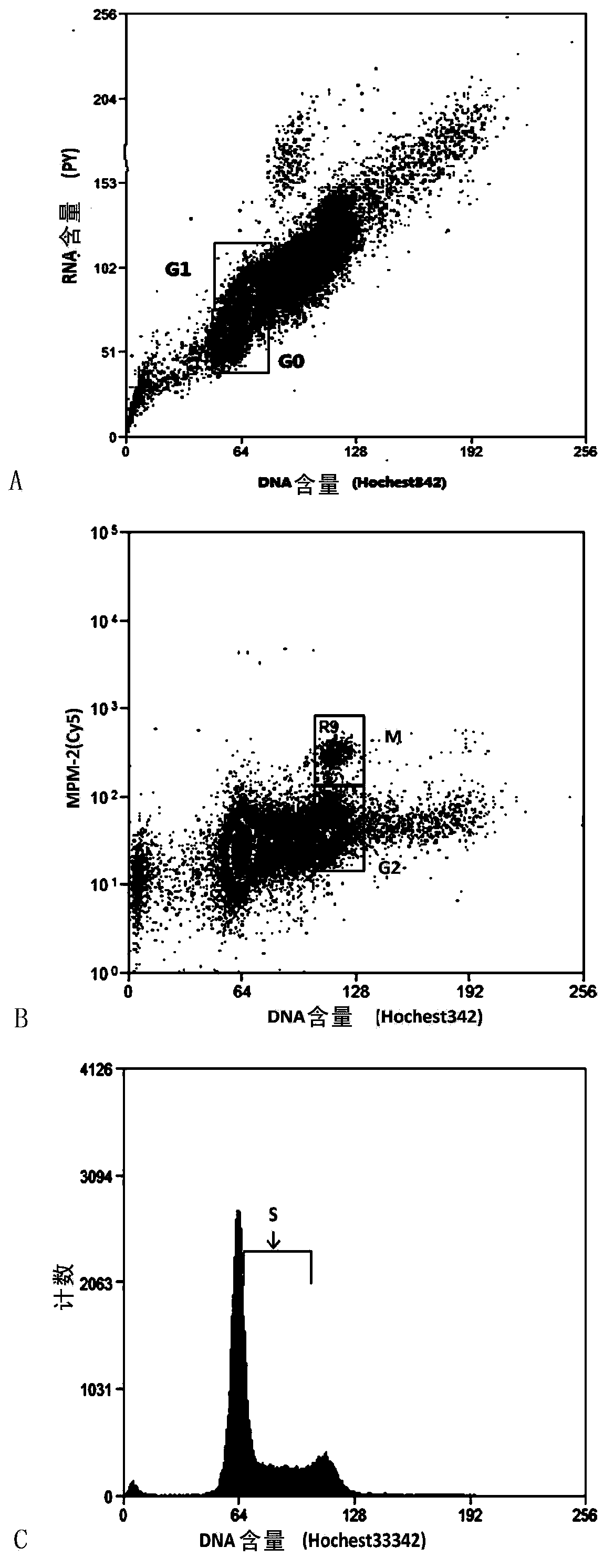
The invention relates to a method for accurately distinguishing cell cycle. According to change rules of DNA and RNA in cell nucleus in cell cycle and the characteristic of protein phosphorylation in cell nucleus in mitotic phase, through cell localization by multiple parameters and by fluorescence detection with a flow cytometer, the cell cycle is effectively and accurately classified into five groups, namely G0, G1, S, G2 and M. The invention discloses, for the first time, a method for accurately distinguishing cell cycle. The method is simple and easy to operate. By the method, the defect that cell cycle cannot be distinguished accurately by a present method is solved. Results of the method provided by the invention are stable and reliable, and the method is suitable for detection of various cells.

Description

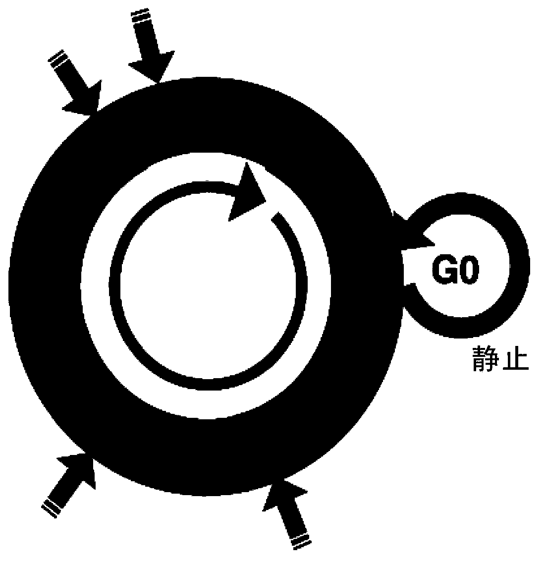
technical field [0001] The invention belongs to the field of biotechnology, and more specifically, the invention relates to an analysis method for accurately distinguishing cell cycle. Background technique [0002] The cell cycle refers to the process that cells that proliferate in mitosis go through from the end of parental division to the end of daughter cell division. During this process, the genetic material of the cell is replicated and doubled, and is equally distributed to the two daughter cells at the end of division. Usually the cell cycle consists of G0 phase, G1 phase, S phase, G2 phase and M phase, such as figure 1 . Cells are in a static state in the G0 phase; G1 phase: the cell begins to synthesize RNA and protein, but the DNA content remains diploid; S phase: DNA synthesis begins, and the DNA content in the nucleus is between the G1 phase and the During the G2 phase, when the DNA replication finishes and becomes quadruploid, the cell enters the G2 phase. Ce...

Claims


Application Information

Patent Timeline
IPC IPC(8): G01N15/14
Inventor 邱琳陈峰
Owner SHANGHAI INST OF BIOLOGICAL SCI CHINESE ACAD OF SCI
Why Eureka
Try Eureka