
Pectoralis segmentation method in breast image

A technology for image center and pectoralis, which is applied in the field of medical image processing, can solve the problems that the threshold cannot be adjusted in real time, the pectoralis cannot be effectively segmented, and the accuracy of the segmentation result is low, so as to suppress large-scale overflow, high accuracy, and improve robustness Effect

Active Publication Date: 2014-12-03
SHANGHAI UNITED IMAGING HEALTHCARE
View PDF4 Cites 7 Cited by
  • Summary
  • Abstract
  • Description
  • Claims
  • Application Information

AI Technical Summary

Problems solved by technology

[0005] The problem solved by the present invention is to provide a method for segmenting pectoral muscles in breast images, which is used to solve the problem that the method for segmenting pectoral muscles in MLO breast images cannot effectively segment irregular or blurred pectoral muscles, and the threshold cannot be adjusted in real time, and only empirical values ​​can be used. , the selection of parameters is greatly affected by noise, the segmentation result is less accurate, and the robustness is poor

Method used


Image

  • Pectoralis segmentation method in breast image
  • Pectoralis segmentation method in breast image
  • Pectoralis segmentation method in breast image

Examples


Embodiment Construction

[0063] In the following description, numerous specific details are set forth in order to provide a thorough understanding of the present invention. However, the present invention can be implemented in many other ways different from those described here, and those skilled in the art can make similar extensions without violating the connotation of the present invention, so the present invention is not limited by the specific implementations disclosed below.

[0064] Secondly, the present invention is described in detail by means of schematic diagrams. When describing the embodiments of the present invention in detail, for convenience of explanation, the schematic diagrams are only examples, which should not limit the protection scope of the present invention.

[0065] In order to solve the technical problems in the background technology, the present invention provides a method for segmenting pectoralis in mammary gland images. Firstly, a morphologically restricted region growth m...


PUM


Abstract

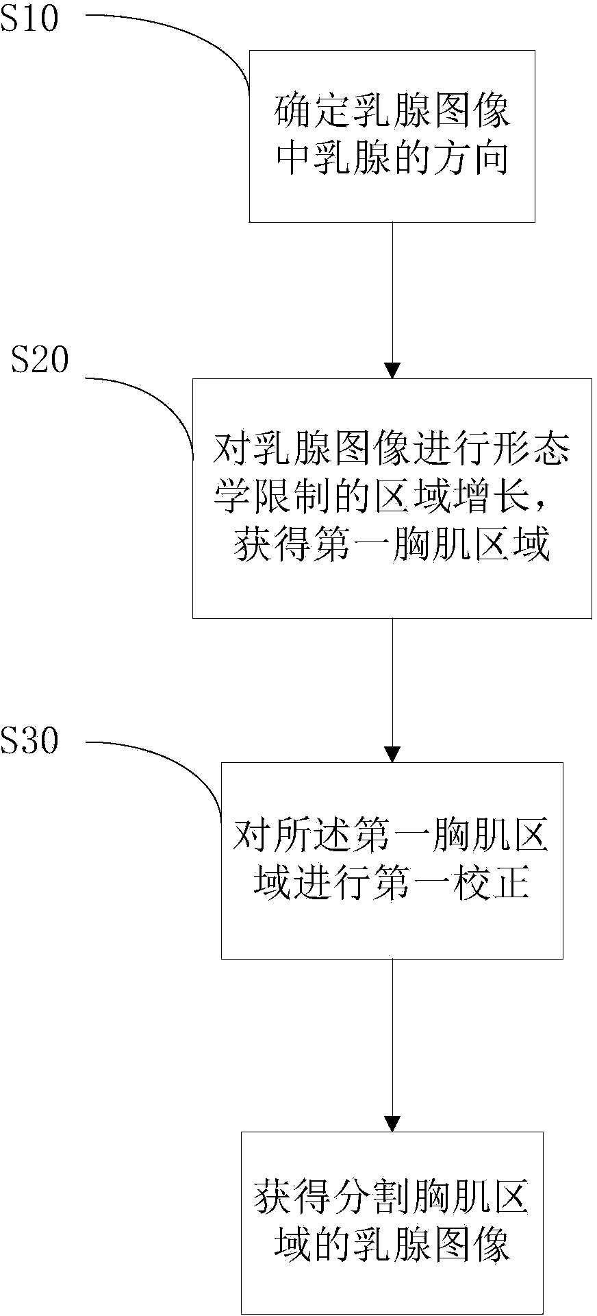
The invention provides a pectoralis segmentation method in a breast image. The pectoralis segmentation method in the breast image comprises the following steps: carrying out morphological restriction region growing to a breast image to obtain a first pectoralis area; and carrying out first correction to the first pectoralis area to obtain the breast image of a pectoralis segmentation area. The pectoralis segmentation method provided by the invention has an automatic correction function, pectoralis parts can be quickly and effectively segmented and removed in a mediolateral oblique position mammogram, and the pectoralis segmentation method exhibits high accuracy and robustness. For multi-level greyscales, the pectoralis with fuzzy edges and irregular edges can be effectively segmented.

Description

technical field [0001] The invention relates to the field of medical image processing, in particular to a method for segmenting pectoral muscles in breast images. Background technique [0002] Removing the human pectoralis region in the medial lateral oblique (MLO) position is a basic step in the preprocessing of mammograms (Mammograms) computer-aided diagnosis (Computer-aided Diagnosis, CAD) system. In the breast image at the MLO position, the pectoralis region is usually located in the upper left corner or upper right corner of the image, and generally has a gentle edge, a triangular shape, and the highest or higher brightness in the entire image. Removing the pectoral muscle step of the MLO image can avoid adverse effects on subsequent processing, such as image regularization in tumor segmentation, breast density estimation, etc., and can effectively suppress false positives for breast CAD, thereby improving the detection rate of lesions. [0003] In the prior art, metho...

Claims


Application Information

Patent Timeline
Patent Type & Authority Applications(China)
IPC IPC(8): G06T7/00
Inventor 李华石海林王瀟崧
Owner SHANGHAI UNITED IMAGING HEALTHCARE
Learn More