Atlas-free brain tissue segmentation method

A separation method and image segmentation technology, applied in image analysis, image data processing, medical science, etc., can solve problems such as degradation of segmentation results, massive calculations, and inaccurate brains

Inactive Publication Date: 2014-10-15
SIEMENS HEALTHCARE GMBH
View PDF4 Cites 4 Cited by
  • Summary
  • Abstract
  • Description
  • Claims
  • Application Information

AI Technical Summary

Problems solved by technology

The applied existing atlas information may thus lead to degradation of the segmentation results and is not precise enough to be applied to each specific brain
In addition, it requires a lot of calculation

Method used


Image

  • Atlas-free brain tissue segmentation method
  • Atlas-free brain tissue segmentation method

Examples


Embodiment Construction

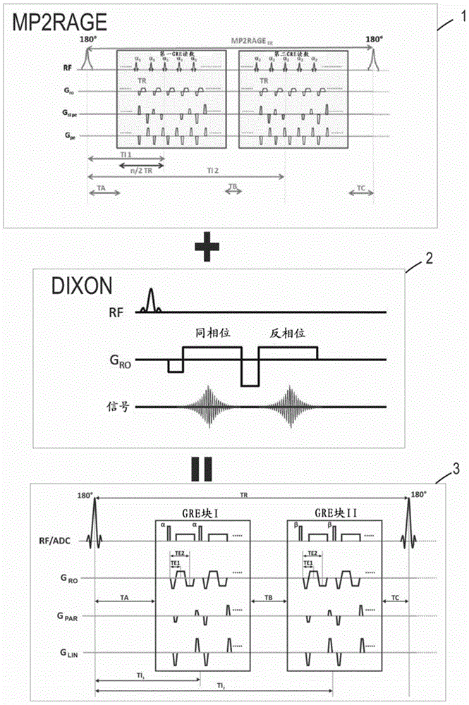
[0041] figure 1 and figure 2 A preferred embodiment of the non-spectral magnetic resonance imaging method according to the invention is schematically shown. The method is configured to image at least a portion of the brain and includes the steps of:

[0042] Using an MRI sequence 3, the MRI sequence 3 is configured to acquire two image volumes I1, I2, I1', I2' of the portion, respectively for each echo at different inversion times TI1, TI2 within a single acquisition First image volume I1, I1' and second image volume I2, I2' at time TE1, TE2, where the MRI sequence 3 is a double-echo MP2RAGE using the Dixon method 2 to acquire the fat-water separation image of the portion Sequence 1, said MP2RAGE sequence 1 is preferably performed using GeneRalized Autocalibrating Partially Parallel Acquisition (GRAPPA) and is specifically characterized by the following parameters:

[0043] TI1 / TI2 / TR=700 / 2500 / 5000ms,

[0044] TE1 / TE2=2.44 / 6.06ms,

[0045] GRAPPA has a reduction factor R...


PUM


Abstract

The invention relates to an atlas-free brain tissue segmentation method. An MRI sequence configured for acquiring two image volumes of the part at different inversion times within a single acquisition is combined to a fat-water separation method for acquiring a fat-water separated image. For each echo time two image volumes are acquired, respectively a first image volume and a second image volume at the first echo time, and a first image volume and a second image volume at the second echo time, and combined to a uniform image. The acquired images are combined to form a final uniform image, a final fat-water separated image, and a final second image volume that are fed into a multichannel image segmentation algorithm using a Markov random field model for segmenting the part into multiple classes of cranial tissues, in order to obtain a segmented image of said part.

Description

technical field [0001] The present invention relates to methods and systems for removing non-brain tissue in magnetic resonance (MR) imaging. In particular, the present invention relates to methods and systems for removing image portions representing non-brain tissue such as skull, scalp, blood vessels or meninges from MR neuroimages, and for automatic segmentation of brain magnetic resonance images. Background technique [0002] In order to acquire images using MR imaging techniques, the object to be imaged is placed in a strong static magnetic field B0 which forces the magnetic moments of the hydrogen nuclei associated with the spins of the object's hydrogen nuclei to assume parallel or opposite directions with respect to the static magnetic field parallel orientation. The spin axis is not perfectly aligned with the static magnetic field, but precesses at a characteristic frequency about the direction axis of the static magnetic field, resulting in a net magnetization in ...

Claims


Application Information

Patent Timeline
Patent Type & Authority Applications(China)
IPC IPC(8): G06T7/00A61B5/055
CPCG01R33/5608G01R33/4828
Inventor 亚历克西斯·罗什托比亚斯·科伯贡纳尔·克吕格尔
Owner SIEMENS HEALTHCARE GMBH
Why Eureka
Try Eureka