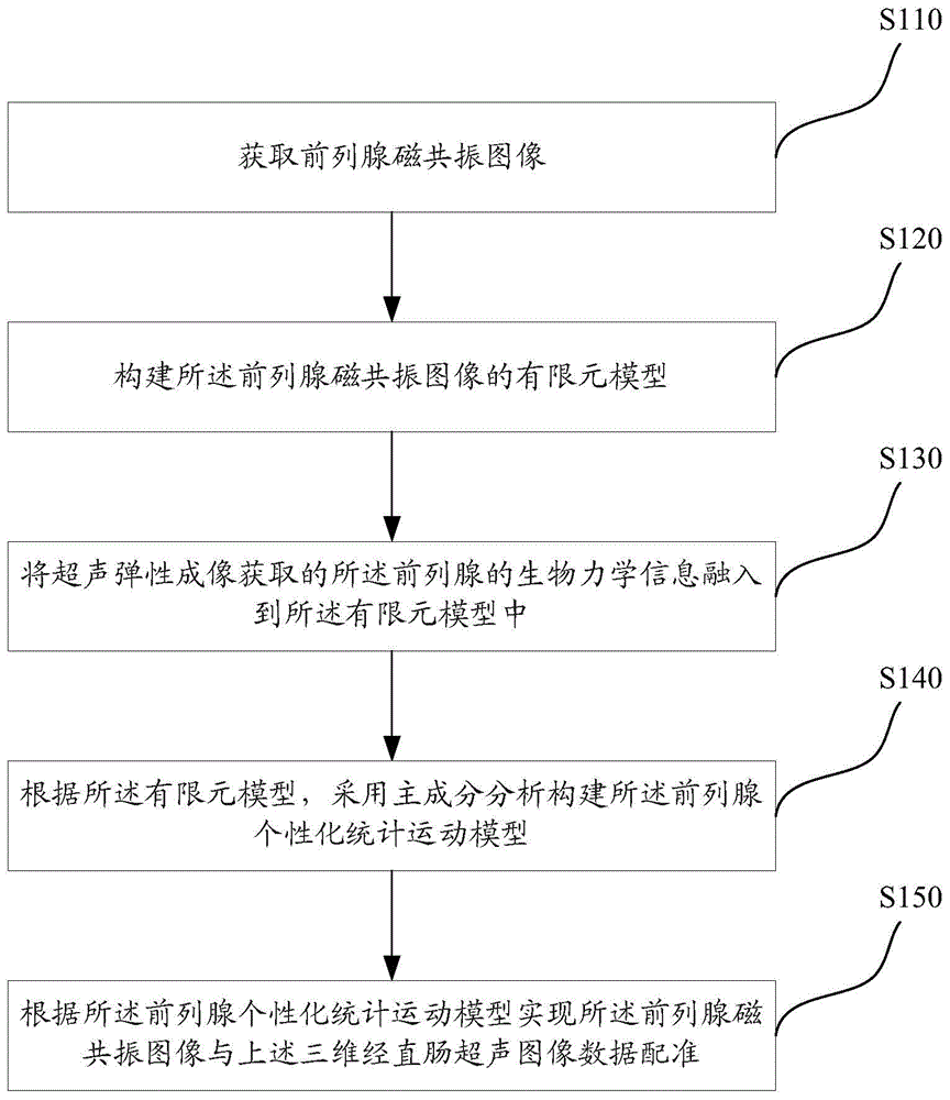
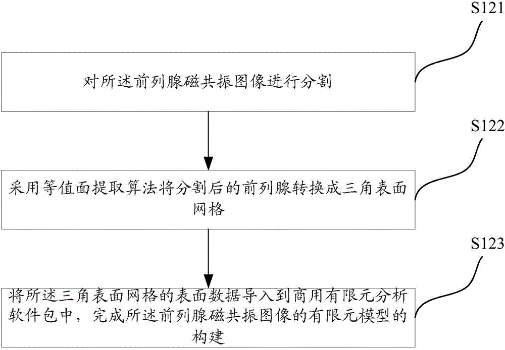
Ultrasound and magnetic resonance image fusion and registration method
A magnetic resonance image and registration technology, applied in the field of medical image processing, can solve the problems of low accuracy and lack of clinical application, etc., and achieve the effect of accurate finite element model and accurate data registration
- Summary
- Abstract
- Description
- Claims
- Application Information
AI Technical Summary
Problems solved by technology
Method used
Image

Examples
Embodiment
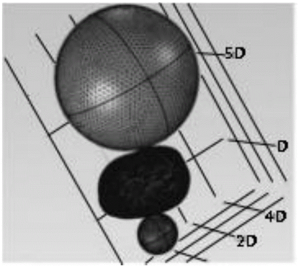
[0085] In order to verify the accuracy and feasibility of the method, we used two different commercial prostate phantoms 053-MM and 053-AEF (CIRS, Norfolk, USA) in the experiment. Magnetic resonance imaging measurements were performed using a 3.0T magnetic resonance scanner with 32-channel head coils. 3D TRUS and ultrasound elastography images were acquired from DC-8 (Mindray, ShenZhen, China) and Aixplorer (Supersonic, France) ultrasound systems, respectively. see Figure 7 , are images of phantom 053-MM in different imaging modes, where (a) is phantom 053-MM, (b) is the cross-section of MRI-T2 image, (c) is B-mode ultrasound, (d) for ultrasound elastography.
[0086] In the experiments, prostate PMSSs were constructed separately for these two different prostate phantoms. Based on elastography, Young's modulus (E=30 kPa in 053-MM, E=20 kPa in 053-AEF) was assigned to the prostate model. To demonstrate the merits of our personalization approach, we construct a generalized ...
PUM
 Abstract
Description
Claims
Application Information
 - R&D
- Intellectual Property
- Life Sciences
- Materials
- Tech Scout
- Unparalleled Data Quality
- Higher Quality Content
- 60% Fewer Hallucinations
Browse by: Latest US Patents, China's latest patents, Technical Efficacy Thesaurus, Application Domain, Technology Topic, Popular Technical Reports.
© 2025 PatSnap. All rights reserved.Legal|Privacy policy|Modern Slavery Act Transparency Statement|Sitemap|About US| Contact US: help@patsnap.com