
Method for detecting classical swine fever virus specific antibody in pig saliva

A swine fever virus and specific technology, applied in the biological field, can solve the problems of small sample size, low proportion of pig herds, and high animal stress, and achieve convenient transportation and storage, save sampling time, and strong resistance. Effect

Inactive Publication Date: 2016-08-17
杭州贝尔塔生物技术有限公司
View PDF7 Cites 0 Cited by
  • Summary
  • Abstract
  • Description
  • Claims
  • Application Information

AI Technical Summary

Problems solved by technology

However, there are many limitations in this sampling method. On the one hand, this method of collecting blood and separating serum is time-consuming and laborious. The number of samples collected is not large, and the proportion of the pig population is not high, and the stress on animals is very large.

Method used


Image

  • Method for detecting classical swine fever virus specific antibody in pig saliva
  • Method for detecting classical swine fever virus specific antibody in pig saliva
  • Method for detecting classical swine fever virus specific antibody in pig saliva

Examples


preparation example Construction

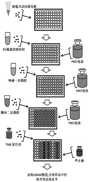
[0022] 1) Preparation, inactivation and antigen-coating of 96-well polystyrene reaction plate for whole swine fever virus antigen;

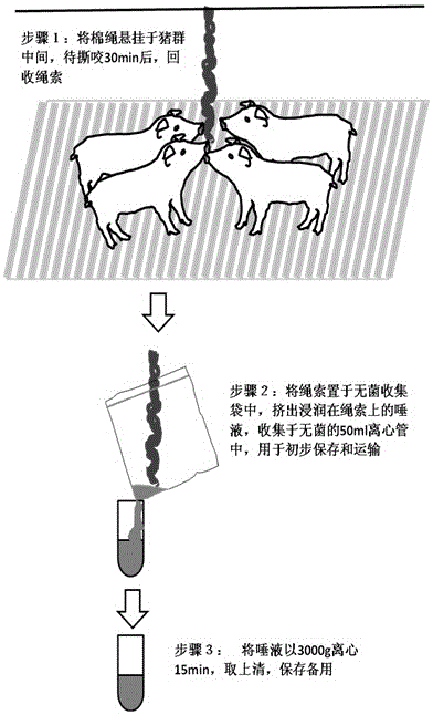
[0023] 2) Use the hanging cotton rope method to induce animals to chew, collect saliva, obtain the primary antibody of saliva after processing, and save it for later use;

[0024] 3) After diluting the saliva primary antibody 1:1 with PBST solution, add it to the reaction wells of the antigen-coated 96-well polystyrene reaction plate according to the number, add 100ul to each reaction well, and react at 22-28°C with saturated humidity for 1 hours, wash and set aside;

[0025] 4) Dilute horseradish peroxidase-labeled goat anti-pig IgG-FC antibody at 1:2000 with PBST solution, and add it to the reaction wells of a 96-well polystyrene reaction plate in sequence, adding 100ul to each reaction well , 22-28 ℃ saturated humidity and dark reaction for 30 minutes, washed with PBST for later use;

[0026] 5) After mixing TMB Chromogenic Agent A and B at ...

Embodiment

[0032] 1) Infect ST cells with the commercialized CSFV vaccine C strain at a concentration of 1000 TCID50 / ml (half cell infection dose / ml). After culturing for 48 hours, collect the virus liquid, centrifuge at 4000g for 10min, and take the supernatant; centrifuge at 35000g After 1 h, the purified virus was obtained and the TCID50 was titrated. Put the virus liquid in a 65°C water bath and inactivate it for 30 minutes to obtain the purified inactivated antigen of the whole swine fever virus. Dilute the inactivated antigen to 10000 TCID50 / ml with pH 9.6 carbonate buffer, coat 96-well polystyrene ELISA reaction plate at a concentration of 100ul / well, and incubate at 22-28°C for 1h under saturated humidity, then Turn to 4°C and incubate for 16h. Then discard the coating solution, pat dry the ELISA plate, add 5% skimmed milk powder diluted with PBST (pH7.3, 0.05% tween-20) 100ul / well, and block at 22-28°C for 2h. Discard the blocking solution, pat dry the ELISA plate, add 150 ul ...


PUM


Abstract

The present invention discloses a method for detecting classical swine fever virus specific antibody in pig saliva. The method comprises: 1) preparing classical swine fever totivirus antigen, inactivating, and coating a 96-well polystyrene reaction plate; 2) adopting a cotton rope suspending method, collecting saliva, and treating to obtain a saliva first antibody; 3) adding the saliva first antibody to an antigen coated reaction plate according to the number, carrying out a 22-28 DEG C saturated humidity reaction for 1 h, and washing; 4) sequentially adding horseradish peroxidase-labeled goat anti-pig IgG-FC antibody to the reaction plate, carrying out a 22-28 DEG C saturated humidity reaction for 30 min under a dark condition, and washing; 5) mixing TMB coloring agents solution A and solution B according to a ratio of 1:1, sequentially adding to the reaction plate, carrying out a reaction under a dark condition for 15 min, and adding a termination solution; and 6) placing into an enzyme-labeling measuring instrument, reading the OD650 data, and determining the result. According to the present invention, the problem of single animal blood collection is avoided, and the method is suitable for large scale investigation of classical swine fever antibody in pig herd.

Description

technical field [0001] The invention relates to the field of biotechnology, in particular to a method for detecting the specific antibody of swine fever virus in swine saliva. Background technique [0002] Classical swine fever (CSF) is currently the most important disease that endangers the pig industry in my country. Its pathological features are degeneration of small blood vessel walls, resulting in multiple hemorrhage, infarction and necrosis of internal organs. The main features are hemorrhage and fever. , is acute or chronic, and is extremely harmful to the pig industry. [0003] Classical swine fever virus (CSFV) is the pathogen that causes swine fever. The disease has the characteristics of high contact and lethality, rapid transmission, widespread prevalence, high morbidity and mortality. Clinically, the disease can be divided into acute, subacute, chronic, atypical and non-obvious types. Acute CSF is induced by virulent strains and generally results in high morbid...

Claims


Application Information

Patent Timeline
Patent Type & Authority Patents(China)
IPC IPC(8): G01N33/569
CPCG01N33/54373G01N33/56983
Inventor 李龙章叶恩曹洋洋
Owner 杭州贝尔塔生物技术有限公司
Learn More