
Non-invasive diagnostic device for gastrointestinal precancerous lesions

A technology for precancerous lesions and gastrointestinal tract, which is used in diagnosis, in vivo radio detectors, medical science, etc. It can solve the problems of manual intervention, inability to meet the needs of diagnosis of diseases of the whole digestive tract, and the existence of blind spots in diagnosis, and achieve automatic control effect

Active Publication Date: 2015-10-28
SHANGHAI JIAOTONG UNIV
View PDF10 Cites 0 Cited by
  • Summary
  • Abstract
  • Description
  • Claims
  • Application Information

AI Technical Summary

Problems solved by technology

However, this technology is also based on traditional endoscopy. Although it has new technical connotations, it also faces the defects of manual intervention, pain in diagnosis and treatment, and blind spots in diagnosis, which cannot meet the clinical needs of diagnosing diseases of the entire digestive tract.

Method used


Image

  • Non-invasive diagnostic device for gastrointestinal precancerous lesions
  • Non-invasive diagnostic device for gastrointestinal precancerous lesions
  • Non-invasive diagnostic device for gastrointestinal precancerous lesions

Examples


Embodiment 1

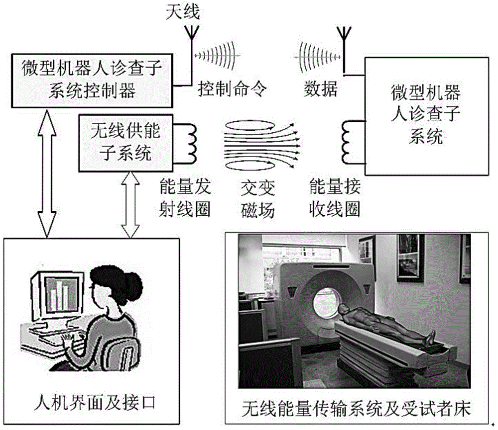
[0028] Such as figure 1 As shown, the non-invasive diagnosis system for gastrointestinal precancerous lesions in this embodiment includes: white light / fluorescence image acquisition and wireless transmission micro-robot diagnosis subsystem in the cavity of the gastrointestinal tract, lying bed and driving subsystem, man-machine interface and Control subsystem, wireless energy supply subsystem.

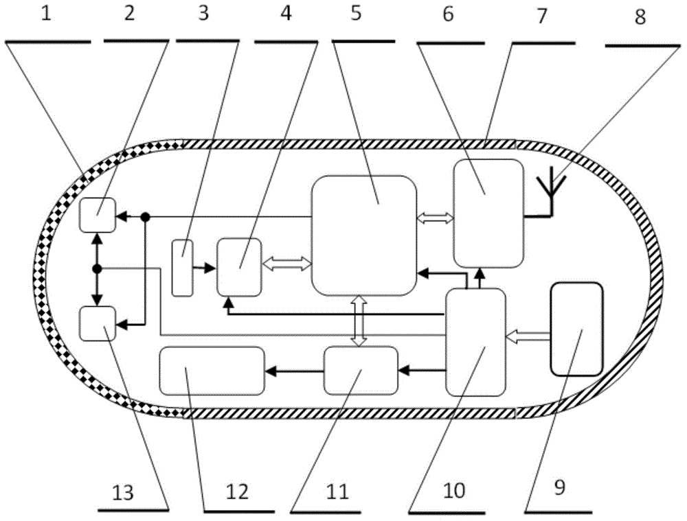
[0029] Such as figure 2 As shown, the white light / fluorescence image acquisition and wireless transmission micro-robot diagnosis subsystem in the gastrointestinal tract cavity of this embodiment includes: a medical transparent hemispherical shell 1, an ultraviolet monochromatic light source 2, a short-focus lens 3, an imaging device 4, Microprocessor 5, wireless communication module 6, medical housing 7, transceiver antenna 8, wireless energy receiving module 9, power management module 10, micro-robot drive control module 11, micro-robot walking mechanism 12, white light source 13, w...


PUM


Abstract

The invention provides a noninvasive examination device for gastrointestinal precancerous lesions. The noninvasive examination device comprises a gastrointestinal intracavity white light / fluorescence image acquisition and wireless transmission micro-robot examination subsystem, a lying-in-bed driving subsystem, a human-computer interface control subsystem and a wireless energy supply subsystem. Through a wireless energy supply and active control system, the noninvasive examination device can be controllable in a gastrointestinal tract of a testee and can be used for examination for a long time, examination results can be sent outside the body of the testee in a wireless mode, and consequently noninvasive and painless examination of the whole digestive tract can be achieved.

Description

technical field [0001] The invention relates to a device in the technical field of non-invasive medical devices, in particular to a device for non-invasive diagnosis of precancerous lesions of the gastrointestinal tract. Background technique [0002] Gastrointestinal cancer has been the malignant tumor with the highest morbidity and mortality for a long time. In my country, the number of new cases of bowel cancer (including colon cancer and rectal cancer) is about 130,000 to 160,000, and the number of deaths is about 60,000 to 90,000, with an increasing trend year by year. Gastrointestinal cancer is hidden, and many patients have reached the advanced stage when they see a doctor, which delays the best time for treatment. Clinical practice shows that the survival rate of early gastrointestinal cancers after good and timely treatment for 5 years and above is over 90%, and some can even be cured. Therefore, improving the diagnosis level of early gastrointestinal cancer is par...

Claims


Application Information

Patent Timeline
Patent Type & Authority Patents(China)
IPC IPC(8): A61B1/00A61B5/07A61B19/00
Inventor 颜国正刘刚王志武刘大生徐文铭朱柄全曾堃虞震芬
Owner SHANGHAI JIAOTONG UNIV
Learn More