
A kind of pathological automatic dna staining system and staining method

A dyeing system and pathological technology, applied in the field of pathological detection, can solve problems such as skin exposure to strong corrosive gases, pathological technicians’ health threats, unfavorable reuse of dye solution, etc., to achieve excellent anti-volatility, fast computing speed, and convenience The effect of calling at any time

Active Publication Date: 2015-10-14
MOTIC XIAMEN MEDICAL DIAGNOSTICS SYST
View PDF8 Cites 0 Cited by
  • Summary
  • Abstract
  • Description
  • Claims
  • Application Information

AI Technical Summary

Problems solved by technology

Almost all known pathological automatic staining machines have problems such as single staining function, poor corrosion resistance, poor washing effect, and poor practicability.
[0004]At the same time, the current Feulgen staining method takes a long time, and the time points of each step need to be specially recorded, which is not conducive to the pathological technician to arrange other work
Manual operation, the dye solution is often not conducive to reuse, and it is easy to expose the skin to strong corrosive gases, which is a great health threat to pathologists

Method used


Image

  • A kind of pathological automatic dna staining system and staining method
  • A kind of pathological automatic dna staining system and staining method
  • A kind of pathological automatic dna staining system and staining method

Examples


Embodiment 1

[0070] Figure 10 It is the staining effect diagram of pig liver smear completed by the staining method and program parameter setting at 35°C shown in Table 1 and Table 2.

[0071] Table 1 35℃ staining method

[0072] serial number

steps

Reagent

cylinder position

time / min

1

fixed

Bohm-Sprenger fixative

2

30

2

rinse

tap water

1

01

3

Acid hydrolysis

5N hydrochloric acid

3

25

4

rinse

tap water

1

01

5

to dye

Thionin stain

4

40

[0073] 6

rinse

tap water

1

03

7

rinse

distilled water

5

05

8

dehydration

50% ethanol

6

02

9

dehydration

75% ethanol

7

02

10

dehydration

95% ethanol

8

01

11

dehydration

100% ethanol

9

01

12

dehydration

100% ethanol

10

01

[0074] Table 2 35°C d...

Embodiment 2

[0077] Figure 11 It is the dyeing effect diagram of pig liver smear completed by the dyeing method and program parameter setting at 45°C shown in Table 3 and Table 4.

[0078] Table 3 45℃ staining method

[0079] serial number

steps

Reagent

cylinder position

time / min

[0080] 1

fixed

4% paraformaldehyde

2

15

2

rinse

tap water

1

01

3

Acid hydrolysis

3N hydrochloric acid

3

20

4

rinse

tap water

1

01

5

to dye

Schiff reagent

4

25

6

rinse

tap water

1

03

7

rinse

distilled water

5

05

8

dehydration

50% ethanol

6

02

9

dehydration

75% ethanol

7

02

10

dehydration

95% ethanol

8

01

11

dehydration

100% ethanol

9

01

12

dehydration

100% ethanol

10

01

[0081] Table 4 Parameter se...

Embodiment 3

[0084] Figure 12 It is the staining effect diagram of pig liver smear completed by the staining method and program parameter setting at 25°C shown in Table 5 and Table 6.

[0085] Table 5 25℃ staining method

[0086] serial number

[0087] Table 6 Parameter setting table of 25°C dyeing program

[0088] cylinder position

[0089] 5


PUM


Abstract

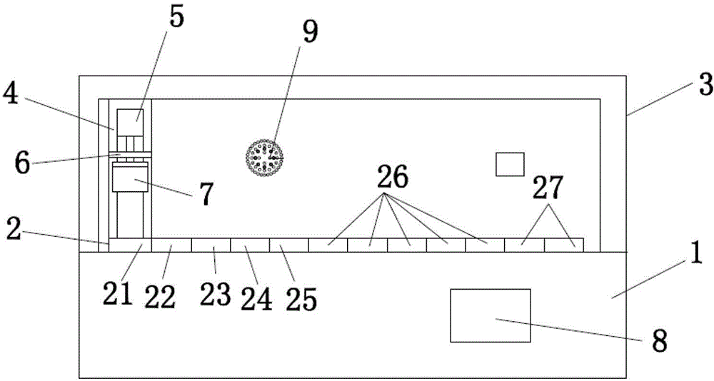
The invention relates to a pathological automatic DNA (deoxyribonucleic acid) staining system. The system comprises a staining platform, a bearing frame, a shifting device and a mechanical arm, wherein a row of liquid tanks are embedded into the staining platform; the bearing frame is vertically connected with the staining platform; the shifting device is connected with the bearing frame in parallel; the mechanical arm is connected with the moving device in a sliding manner, and a mounting device is connected to the mechanical arm; a plurality of staining frames are hung at the lower end of the mounting device; a vent hole is formed in the bearing frame; each device is controlled to work by a microcomputer system, and a control window of the microcomputer system is formed in the side surface of the staining platform; and an air exhaust system and a power-off protection system are arranged in the pathological automatic staining system. By utilizing the technical scheme provided by the invention, the automatic operation of multiple sets of staining methods is realized, the staining is simple and stable, and the number of sheets stained once can be selected according to requirements, thus being suitable for requirements of different users.

Description

technical field [0001] The invention relates to the field of pathological detection, in particular to a device and a staining method for automatic DNA staining of glass slides submitted for pathological inspection. Background technique [0002] In recent years, with the rapid development of computer science, the quantitative analysis technology of pathological images has been widely used in medical diagnosis. Among them, the representative technology of quantitative analysis of cell DNA is of great significance to the early detection of tumors, the prognosis of cancer and the guidance of clinical treatment. The principle is to use Feulgen staining method to stain the nuclear DNA on cell smears, tissue sections or prints, that is, fix with fixative solution, hydrochloric acid acid hydrolysis, stain with Thionin staining solution or Schiff reagent, and pass through the high-precision CCD on the microscope. The camera collects the DNA image of the cell nucleus, and digitally c...

Claims


Application Information

Patent Timeline
IPC IPC(8): G01N1/30G01N1/31
Inventor 唐剑黄荣祥
Owner MOTIC XIAMEN MEDICAL DIAGNOSTICS SYST
Learn More