Embryonic stem cell specific marker GM-CSFR alpha and application thereof
A GM-CSFR and embryonic stem cell technology, applied in the field of embryonic stem cell-specific marker GM-CSFRα, can solve the problem of limited number of embryonic stem cell membrane surface markers, and achieve high specificity and sensitivity
- Summary
- Abstract
- Description
- Claims
- Application Information
AI Technical Summary
Problems solved by technology
Method used
Image

Examples
Embodiment 1
[0019] Example 1 Preparation of Rabbit Monoclonal Antibody
[0020] Using mouse embryonic stem cells (mES) as the immunogen, each time 1×10 8 A mES was used to immunize 3-month-old New Zealand white rabbits for 4 times, with an interval of 3 weeks between each immunization. Serum was collected after four times of immunization, and 1 × 10 8 3 days after boosting, rabbit spleen cells were isolated and fused with myeloma cell 240E-W2 (purchased from Epitomics), and the hybridoma cell line ZJUESRMAB29 (accession number CGMCC) producing GM-CSFRα rabbit monoclonal antibody was screened out. NO.7302), applied to the identification and isolation of stem cells.
Embodiment 2
[0021] Example 2 Application of GM-CSFRα as a marker for embryonic stem cells
[0022] The expression of GM-CSFRα decreases with cell differentiation and is limitedly expressed in adult tissues, which can be used as a new marker for the identification of embryonic stem cells.
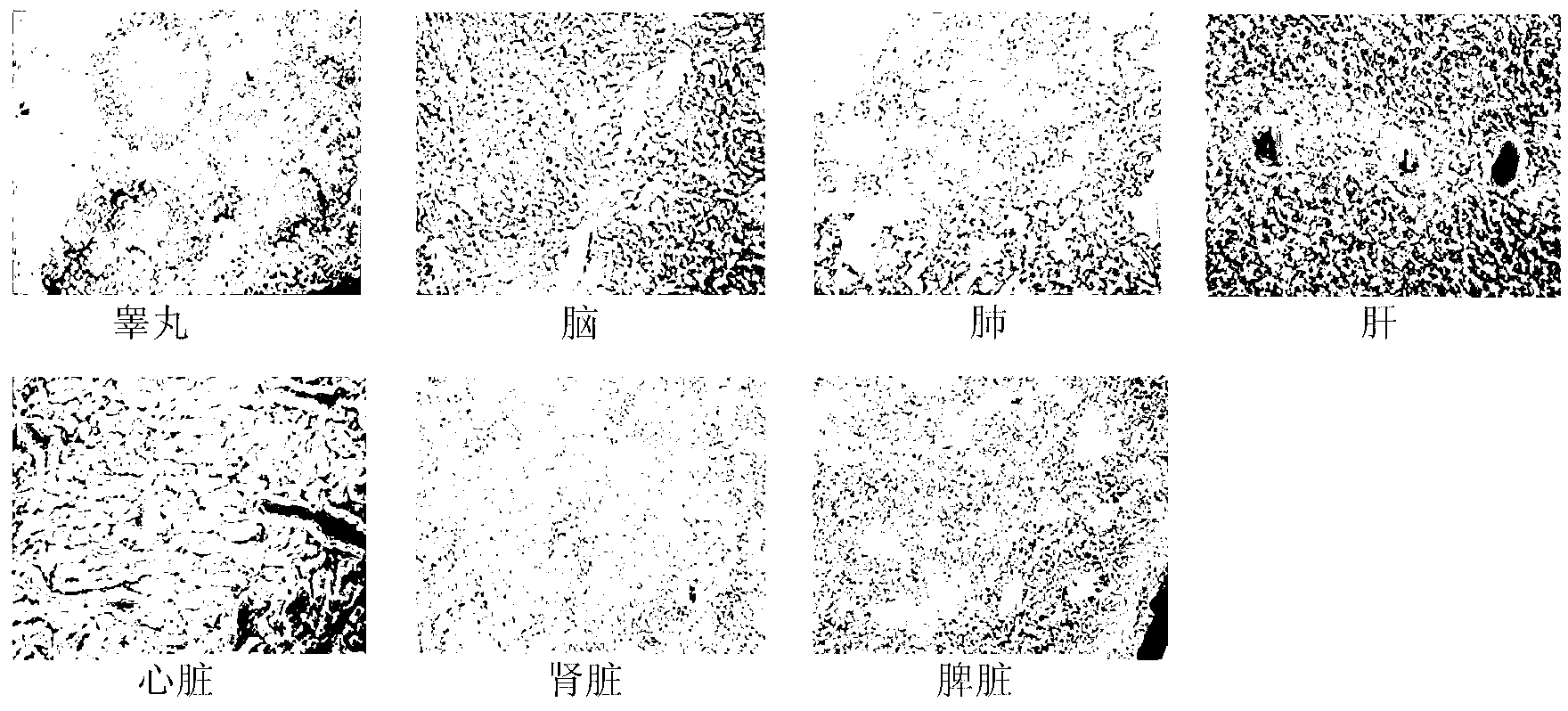
[0023] IHC staining experiment: Prepare the frozen sections of each tissue; place the frozen sections of each tissue in 3% hydrogen peroxide for 10 minutes to inactivate endogenous peroxidase; use blocking solution (containing 10% goat serum and 1% BSA in PBS) at 37°C for 1 hour; add primary antibody (GM-CSFRα rabbit monoclonal antibody prepared in Example 1) and incubate at 37°C for 1 hour; then add goat anti-rabbit-HRP secondary antibody and incubate at 37°C for 1 hour ; Finally, use freshly prepared 3,3'-diaminobenzidine (DAB) to develop color at 37°C for 5 minutes, observe and read the film. The result is as figure 1 shown. The results of IHC staining showed that GM-CSFRα was highly expressed in ...
Embodiment 3
[0024] Example 3 Application of GM-CSFRα as a marker for embryonic stem cells
[0025] ICC staining experiment proved that GM-CSFRα rabbit monoclonal antibody can be applied to the identification and sorting of embryonic stem cells.
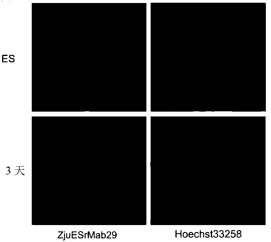
[0026] ICC experiment: Cultured cells were fixed with 4% formaldehyde (Sigma) at 37°C for 1.5 hours; then blocked with blocking solution (PBS containing 10% goat serum and 1% BSA) at 37°C for 1 hour; the blocked cells were added with primary antibody ( The GM-CSFRα rabbit monoclonal antibody prepared in Example 1) was incubated at 37°C for 1 hour; washed with PBS; the washed cells were added with secondary antibody and incubated for 45 minutes at 37°C in the dark; finally stained with 0.5 μg / ml Hochest33258 (Sigma) nuclear 5 minutes; observed and photographed under a fluorescent microscope (Olympus IX-70). The result is as figure 2 shown. GM-CSFRα rabbit monoclonal antibody ICC staining results showed that compared with undifferentiated mES c...
PUM
 Abstract
Description
Claims
Application Information
 - R&D
- Intellectual Property
- Life Sciences
- Materials
- Tech Scout
- Unparalleled Data Quality
- Higher Quality Content
- 60% Fewer Hallucinations
Browse by: Latest US Patents, China's latest patents, Technical Efficacy Thesaurus, Application Domain, Technology Topic, Popular Technical Reports.
© 2025 PatSnap. All rights reserved.Legal|Privacy policy|Modern Slavery Act Transparency Statement|Sitemap|About US| Contact US: help@patsnap.com