
Immunofluorescence staining method for suspension cells

A technique for immunofluorescence staining and suspension cells, which is used in the preparation of test samples to achieve good staining results.

Inactive Publication Date: 2013-06-12
RENJI HOSPITAL AFFILIATED TO SHANGHAI JIAO TONG UNIV SCHOOL OF MEDICINE
View PDF2 Cites 2 Cited by
  • Summary
  • Abstract
  • Description
  • Claims
  • Application Information

AI Technical Summary

Problems solved by technology

However, there is no report on an immunofluorescence staining method for suspension cells

Method used


Image

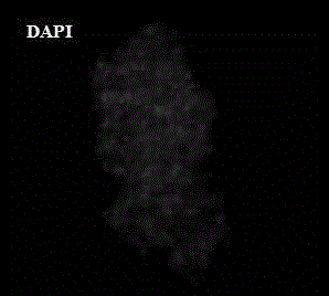
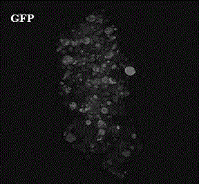
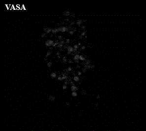
  • Immunofluorescence staining method for suspension cells
  • Immunofluorescence staining method for suspension cells
  • Immunofluorescence staining method for suspension cells

Examples


Embodiment 1

[0032] 1. Experimental materials

[0033] Cells: Mouse iPS cells (induced pluripotent stem cells, induced pluripotent stem cells), cultured in a bacterial culture dish for 5 days to form embryoid bodies (EBs), added 2um of retinoic acid (RA) and cultured for 2 days to induce differentiation EB cells.

[0034] Reagents: 4% paraformaldehyde, phosphate buffer (Hyclone), 1% BSA (Sigma), TritonX-100, primary antibody VASA rabbit anti-mouse (Abcam), fluorescent secondary antibody A11072 goat anti-rabbit (Invitrogen), DAPI, Mounting medium (DAKO).

[0035] Equipment: low-speed centrifuge (Thermo), low-speed shaker, ordinary refrigerator (Midea), laser confocal microscope (Leica).

[0036] 2. Experimental method

[0037] 1. Collect the RA-induced EB cell mass in a 2ml centrifuge tube;

[0038] 2. Add 2ml of PBS, gently invert several times, centrifuge at 800g for 5min, and discard the supernatant;

[0039] 3. Repeat step 2;

[0040] 4. Add 2ml of distilled water, gently invert s...


PUM


Abstract

The invention relates to an immunofluorescence staining method for suspension cells. The immunofluorescence staining method for the suspension cells is characterized by comprising the following steps of: collecting cells into a centrifuge tube; performing 800-gram centrifugation to collect the cells; respectively adding paraformaldehyde to fix, Triton-X100 to pass through and 1 percent bovine serum albumin (BSA) to close; adding a primary antibody and a secondary antibody sequentially to incubate; washing by an 800-gram centrifugation method after the operation of each step; performing diamino phenyl indole (DAPI) staining; and dripping on a glass slide and closing the glass slide to observe. The immunofluorescence staining method for the suspension cells has the advantages that: the immunofluorescence staining process is performed in the centrifuge tube, so the problems that the suspension cells grow on the glass slide difficultly and drop off easily in the test process are solved; the staining result is good; and the method is suitable for the suspension cells and cells which are adhered to the wall difficultly.

Description

technical field [0001] The invention relates to a cell staining method, in particular to an immunofluorescent staining method for suspended cells. Background technique [0002] For traditional cell immunofluorescence staining, methods such as cell climbing or direct smear are usually used to pretreat cells before immunofluorescence staining. For the cells or cell clusters growing in suspension, when the cells are treated by climbing slides, there will often be poor adherence or growth on both sides of the slide at the same time, which will affect the later staining and observation; in addition, in the subsequent staining process , the cells attached to the slides are easy to fall off after repeated washing. [0003] Chinese patent document CN101329230B discloses an improved immunofluorescent cell staining method, including five steps of fixation / permeabilization, permeabilization, surface staining and nuclear staining, washing, and resuspension. The improvement of the stai...

Claims


Application Information

Patent Timeline
Patent Type & Authority Patents(China)
IPC IPC(8): G01N1/30
Inventor 孙璨朱勇杨施胡洪亮李铮
Owner RENJI HOSPITAL AFFILIATED TO SHANGHAI JIAO TONG UNIV SCHOOL OF MEDICINE
Learn More