


Quick Research
Generate reliable direction feasibility study reports for your R&D in just a few steps.

Technical Q&A
Discover and master advanced knowledge NOW. Basics, ideas, possibilities, all at once.

Find Solutions
As an expert in R&D theories, this can generate solutions to your technical problems instantly.

Evaluate Feasibility
Analyze your overall solution with one click, know your potential R&D risks in advance.

Monitor Landscape
Get weekly tech updates, stay abreast of the latest tech innovations and key insights.
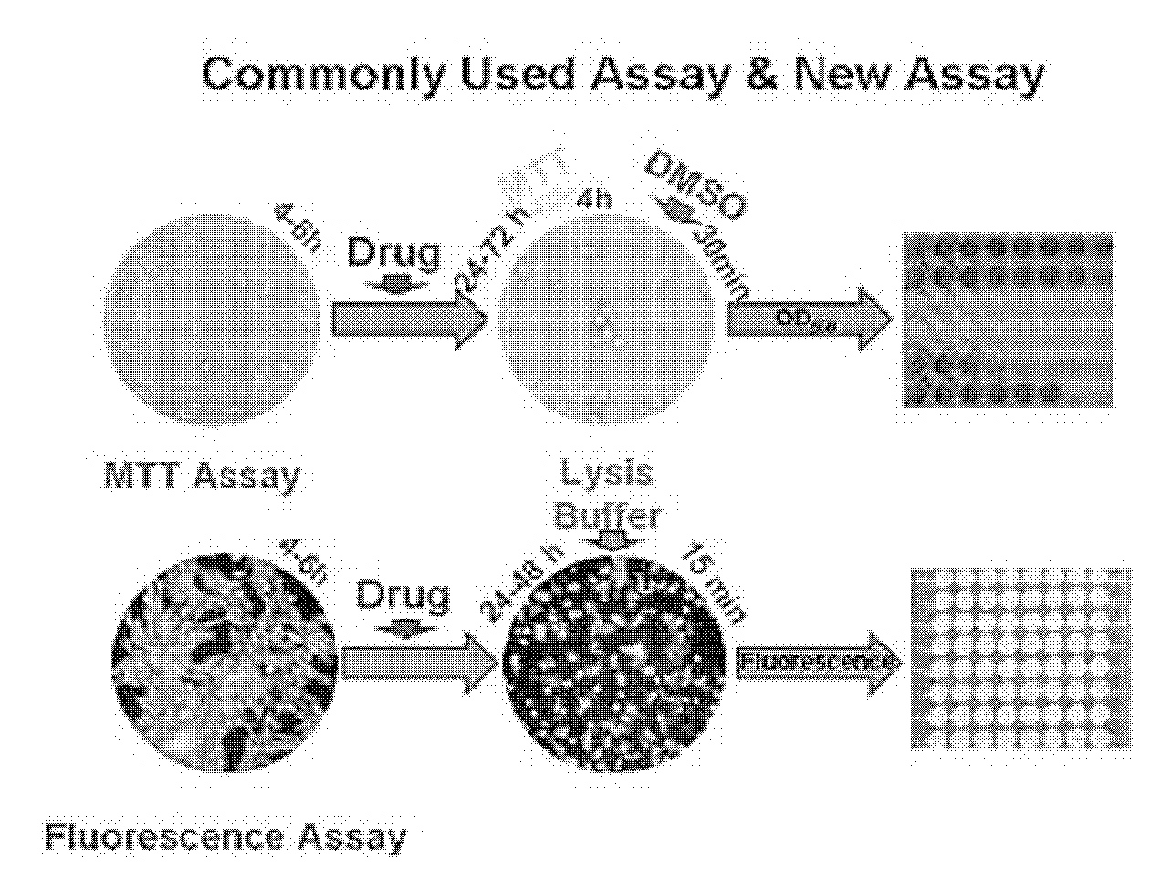
Fluorescent cell model for screening of antitumor drugs, labeling method and application thereof
An anti-tumor drug and cell model technology is applied to a fluorescent cell model for anti-tumor drug screening and its labeling and application fields, and can solve the problems of inability to detect the absolute number of cells, influence on the accuracy of experimental results, and increased workload. To achieve the effect of clear drug action mechanism, small error and easy operation
- Summary
- Abstract
- Description
- Claims
- Application Information
AI Technical Summary
Problems solved by technology
Method used
Image

Examples
Embodiment 1
[0030] Establishment of U2OS cell line (U2OS-EGFP cell line) efficiently and stably expressing enhanced green fluorescent protein (EGFP)
[0031] 1. Transfection of U2OS cells with neo-resistant EGFP plasmid
[0032] 1) U2OS cells were subcultured for 2-3 times in order to maintain the cells in a good growth state and prepare for plating;
[0033] 2) Spread 24-well plates, a 90% full 10cm dish cells can be plated in 24 wells, and spread in 24-well cell culture plates according to the amount of 500 μL medium per well. Observe the cell adhesion every 20 minutes, and shake the plate to spread the cells in the well as evenly as possible;
[0034] 3) Incubate at 37°C for 1-2 hours in 5% CO2, and prepare one well of a 24-well plate for transfection after the cells adhere to the wall;
[0035] 4) Transfection step: according to DNA concentration of 0.4 μg / mL and PEI concentration of 0.8 μg / mL, dissolve in 200 μD DEM basal medium to prepare transfection solution; after incubating at...
Embodiment 2
[0048] Establishment of a method for screening antitumor drugs using U2OS-EGFP cell model
[0049] 1. Screening of anti-tumor drug test scheme by fluorescence analysis method based on U2OS-EGFP cells
[0050] The first stage: use high-concentration samples to analyze the cell growth inhibition rate of the sample, and initially determine whether the sample has the activity of inhibiting cell growth;
[0051] The second stage: In this stage, the samples are divided into high, medium and low concentration groups, and the statistics of the cell growth inhibition rate and the significant difference of the samples are carried out to accurately determine whether the sample has cell growth inhibition activity, and to screen the cells with a cell growth inhibition rate greater than 50%. Samples are hits;
[0052] The third stage: In this stage, the gradient dilution method is used to divide the samples into 9 concentration groups, analyze the relationship between the sample concentrat...
PUM
 Abstract
Description
Claims
Application Information
 - R&D Engineer
- R&D Manager
- IP Professional
- Industry Leading Data Capabilities
- Powerful AI technology
- Patent DNA Extraction
Browse by: Latest US Patents, China's latest patents, Technical Efficacy Thesaurus, Application Domain, Technology Topic, Popular Technical Reports.
© 2024 PatSnap. All rights reserved.Legal|Privacy policy|Modern Slavery Act Transparency Statement|Sitemap|About US| Contact US: help@patsnap.com