
Three-dimensional type-B ultrasound device for realizing conical scanning

A conical scanning, three-dimensional technology, applied in the field of medical devices, can solve problems such as insufficient solution to clinical medical problems, geometric distortion of B-ultrasound images, poor scanning effect, etc., and achieve the effect of controllable scanning range, convenient operation, and stable rotation.

Active Publication Date: 2012-12-19
WENZHOU MEDICAL UNIV
View PDF0 Cites 0 Cited by
  • Summary
  • Abstract
  • Description
  • Claims
  • Application Information

AI Technical Summary

Problems solved by technology

In the prior art, there are two methods of B-ultrasound / UBM with sector scanning. The first one is to use a DC rotating motor to drive the pendulum through a one-stage rotation-swing conversion mechanism, and directly convert the rotational motion into a swing. When the rotation radius of the shaft pin is equal to the swing radius of the pendulum body, the pendulum body can swing at a constant speed, and the scanning head swings according to the cosine law. When there is an error between the rotation radius of the shaft pin and the swing radius of the pendulum body, it will cause Severe geometric distortion in B-ultrasound images
Another method is to use a custom motor to directly drive the pendulum, but its structure is complex, the scanning effect is poor, the accuracy is low, and the cost is high
It can be seen that the sector scanning ultrasonic biomicroscope is not enough to solve the difficult problems of clinical medicine

Method used


Image

  • Three-dimensional type-B ultrasound device for realizing conical scanning
  • Three-dimensional type-B ultrasound device for realizing conical scanning
  • Three-dimensional type-B ultrasound device for realizing conical scanning

Examples


Embodiment Construction

[0017] In order to further explain the technical means and effects that the present invention adopts to achieve the predetermined purpose, the specific implementation method of a three-dimensional B-ultrasound device for conical scanning proposed according to the present invention will be described below in conjunction with the accompanying drawings and preferred embodiments. , structure, feature and effect thereof are described as follows.

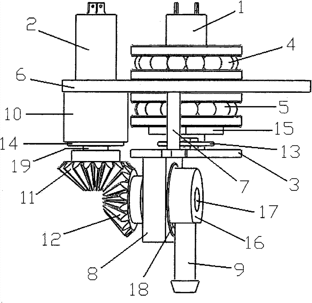
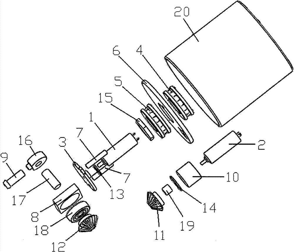
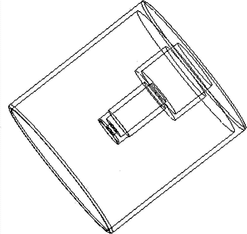
[0018] An embodiment of the present invention provides a three-dimensional B-ultrasound device for conical scanning, such as figure 1 , figure 2 As shown, it includes: housing 20, motor one 1, motor two 2, coupling piece 3, thrust ball bearing one 4, thrust ball bearing two 5, support piece 6, transmission rod 7, bearing fixing piece 8, probe 9, Motor fixing sleeve 10, bevel gear one 12, bevel gear two 11, program controller and control line board (not shown), and described shell 20 is provided with inner and outer two layers, as imag...


PUM


Abstract

The invention discloses a three-dimensional type-B ultrasound device for realizing conical scanning, which is characterized in that: a thrust ball bearing 1, a support sheet, a thrust ball bearing 2 and a nut are sequentially fixed on the outer wall of an inner layer of a shell, a motor 1 and a coupling sheet are connected, a photoelectric encoder 1 is arranged between the nut and the coupling sheet, and two ends of a support rod are respectively and fixedly connected with the coupling sheet and the support sheet; the coupling sheet is fixedly connected with a bearing fixing sheet, a bevel gear 1 and a probe are connected and respectively arranged at two sides of the bearing fixing sheet, and a sensor in the probe is connected with a control circuit board; a motor fixing sleeve is fixed at the periphery of the support sheet, one end of a motor 2 passes through the motor fixing sleeve and is connected with a bevel gear 2, a photoelectric encoder 2 is arranged between the motor 2 and the bevel gear 2, and the bevel gear 2 and the bevel gear 1 are engaged; and the photoelectric encoder 1 and the photoelectric encoder 2 are both connected with the control circuit board which is connected with the program controller. The invention has the advantages of simple structure, convenience of operation, controllable scanning range, simpleness of data process and high scanning efficiency.

Description

[technical field] [0001] The invention relates to the field of medical instruments, in particular to a three-dimensional B-ultrasound device for realizing cone scanning. [Background technique] [0002] The basic principle of B-ultrasound imaging is: emit a group of ultrasonic waves to the human body, scan in a certain direction, and judge the distance and nature of the organs according to the delay time and strength of the echo. After processing by electronic circuits and computers, a B-ultrasound image is formed. Ultrasound Biomicroscope (abbreviation: UBM, English full name: Ultrasound Biomicroscope) is a new type of ophthalmic B-ultrasound imaging detection equipment developed in the early 1990s. UBM uses programmable logic devices to control electronic circuits to excite high-frequency ultrasonic sensors to emit high-frequency ultrasonic waves as a signal source, receive ultrasonic echo signals and perform electronic signal processing to obtain digital images related to...

Claims


Application Information

Patent Timeline
Patent Type & Authority Patents(China)
IPC IPC(8): A61B8/00
Inventor 瞿佳徐亮禹朱明善
Owner WENZHOU MEDICAL UNIV
Learn More