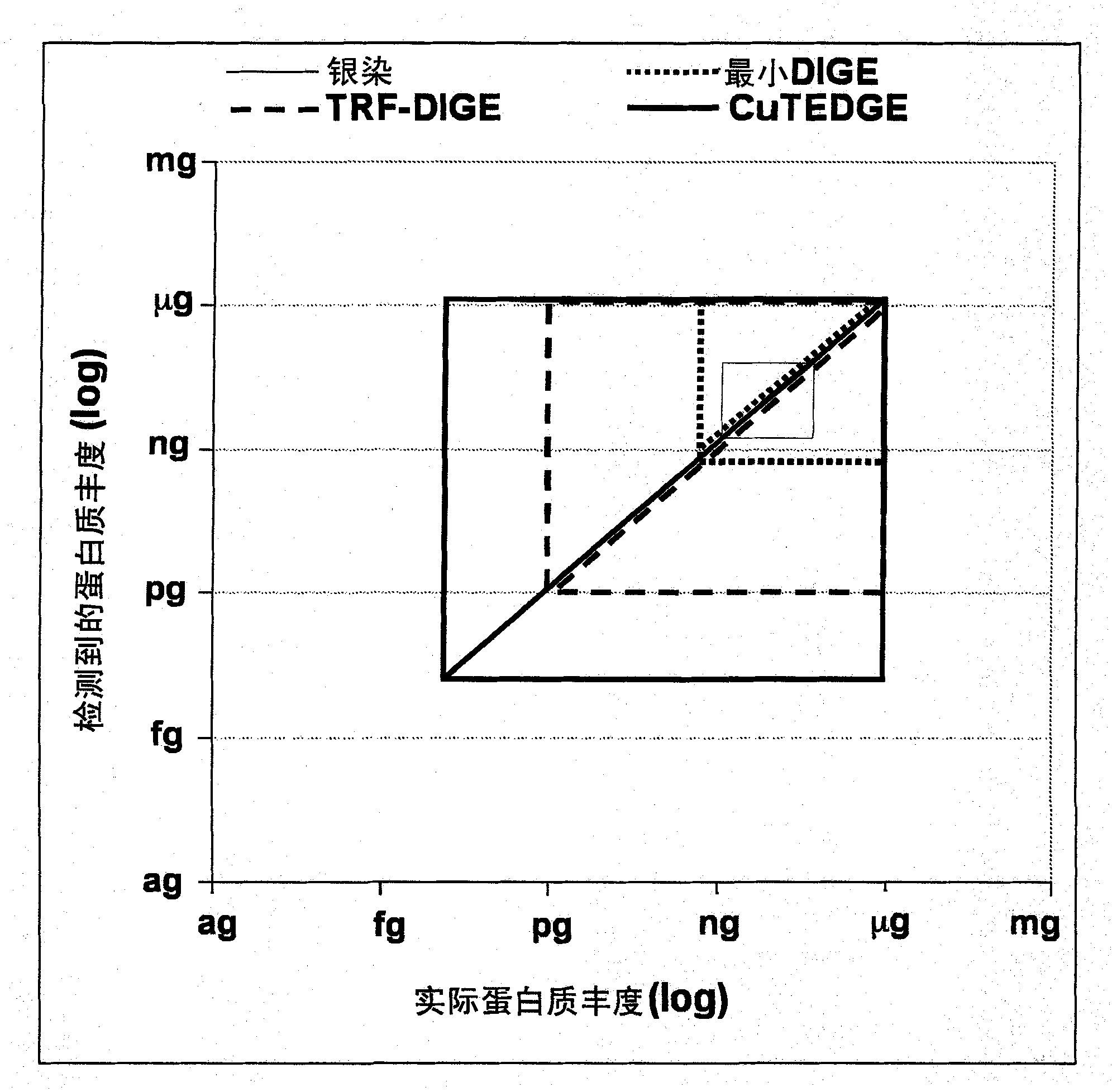
Cumulative time-resolved emission two-dimensional gel electrophoresis
A technology of two-dimensional gel electrophoresis and fluorescent dyes, which is applied to the analysis of materials, material analysis through electromagnetic means, material analysis through optical means, etc., and can solve problems that prevent the use and development of TRF
- Summary
- Abstract
- Description
- Claims
- Application Information
AI Technical Summary
Problems solved by technology
Method used
Image
Examples
Embodiment 1
[0057]In this example, proteins were first extracted from rat airways by lysis lavage (14) in 2-DGE compatible urea-based lysis buffer (7M urea, 2M thiourea, 4% w / v Rapid solubilization of the airway epithelial proteome in CHAPS, 0.5% Triton-X 100, 2% v / v protease inhibitor cocktail). Protein concentrations were determined using the Bradford method, and proteins were separated using 2-DGE. An aliquot of 400 μg protein / sample was diluted to 350 μl with lysis buffer and IPG buffer of the appropriate pH range was added to a final concentration of 1% v / v. By rehydrating overnight at room temperature, protein samples were loaded onto IPG gel strips (GEHealthcare) and passed through the following gradient protocol at 20°C using a Multiphor II Electrophoresis unit electrophoresis instrument and an EPS3501XL power supply electrophoresis tank: 0-50V , 1min.; 50V, 1 hr; 50-1000V, 3 hr; 1000-3500V, 3hr; 3500V, 19hr (total 74.9kVh) for isoelectric focusing (IEF). After IEF, the strips w...
Embodiment 2
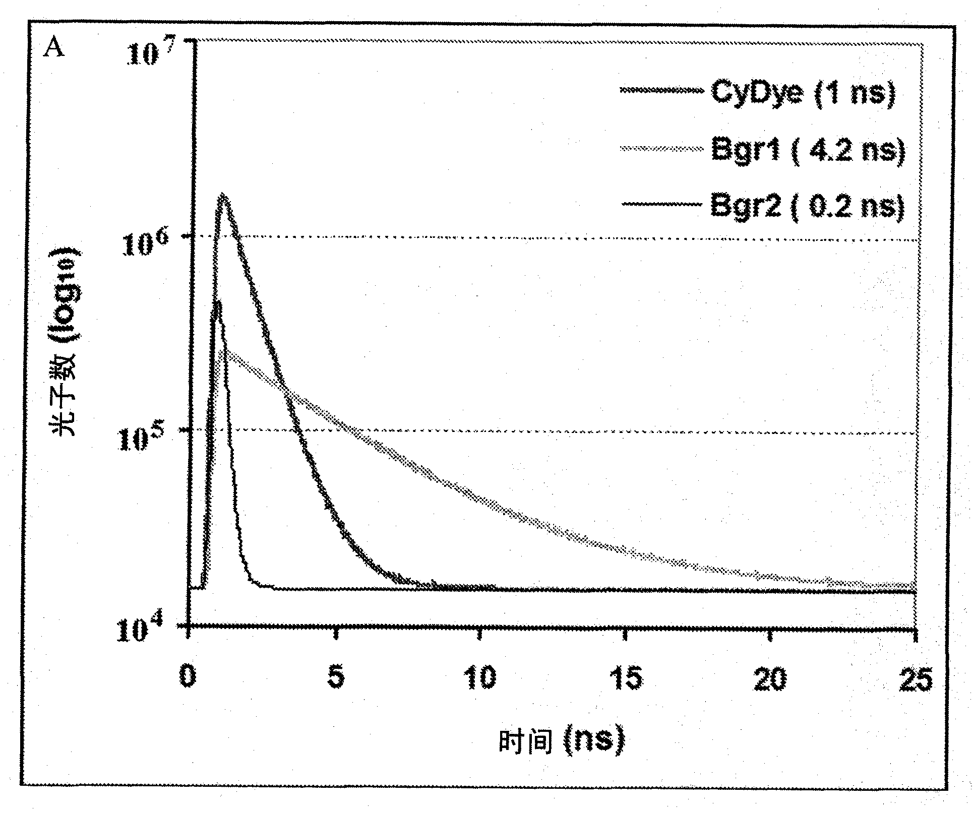
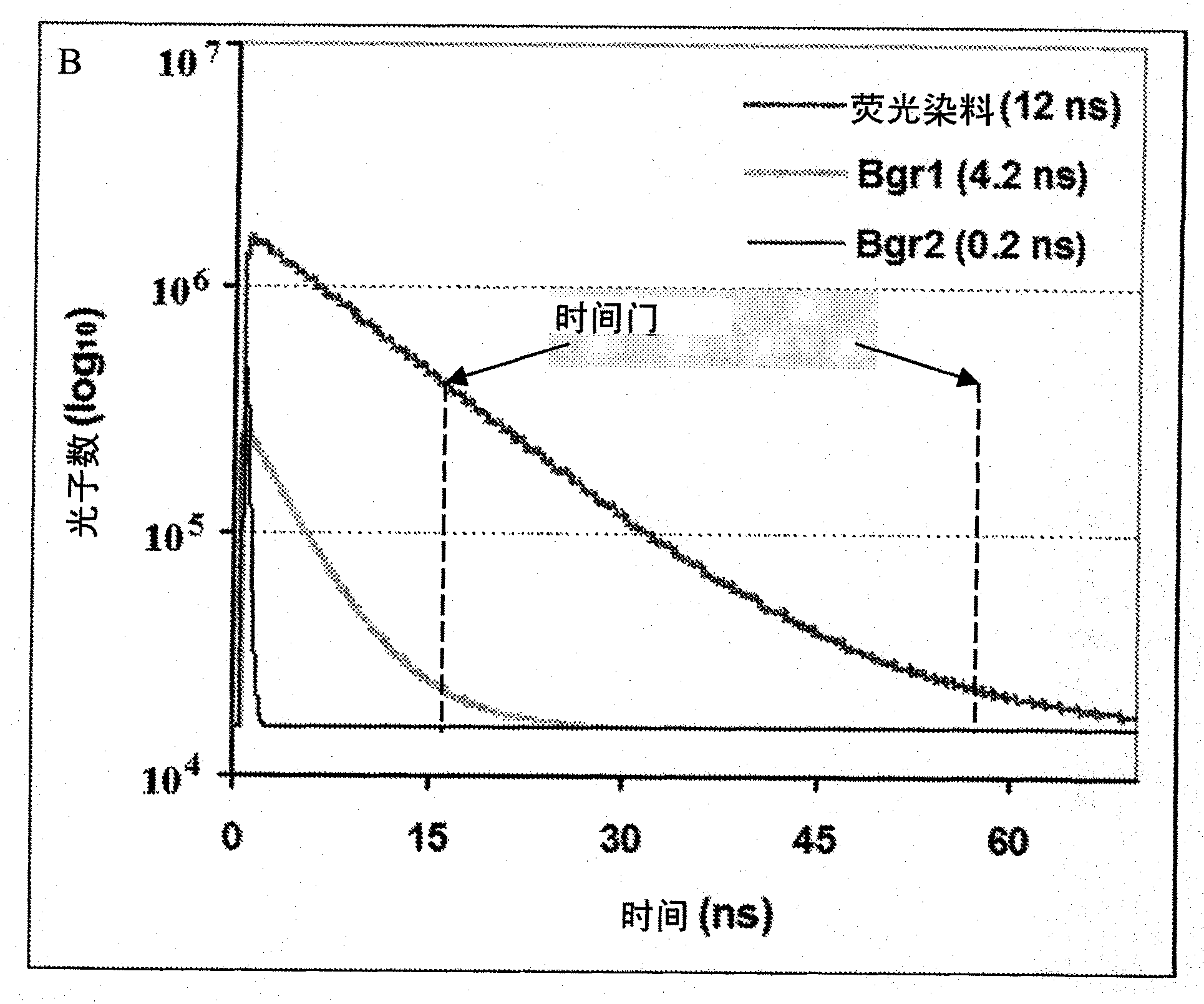
[0063] Macrophages extracted by bronchoalveolar lavage from smoking and never-smoking human subjects were lysed with 2-DGE lysis buffer (see Example 1). Internal standards were generated by pooling equal amounts of protein from all subjects included in the study and were labeled with NHS ester-conjugated Cy2 (minimum DIGE reagent) according to the manufacturer's recommendations (GE Healthcare, Uppsala, Sweden). Protein samples from smoking and never-smoking subjects were randomly divided into two groups and labeled with Cy3 and Cy5 (minimal DIGE reagent), respectively. Cy2-labeled internal standards were co-separated on 2-DGE gels using one Cy3 and one Cy5-labeled sample according to the protocol described in Example 1. Three different protein markers were visualized in an automated sequence of three different scanning protocols using a combination of spectral and time-resolved fluorescence. The specific operation is as follows:
[0064] A) Cy2 fluorescent dye (internal stan...
Embodiment 3
[0069] As an additional step to the protocol described in Example 2, perform SYPRO TM Ruby staining facilitates quantification of total protein content in DIGE gels. In prior art, due to spectral overlap, especially in SYPRO TM Spectral overlap in the emission curves of Ruby, Cy3 and Cy5 makes it impossible to use DIGE and SYPRO in the same 2-DGE gel TM Ruby fluorescent dye. Embodiments of the present invention utilize temporal dimensions to facilitate the incorporation of these methods with standard protocols (as exemplified in FIG. 3 ). After completing the steps outlined in Example 2, following steps B and C outlined in Example 1, in SYPRO TM Total protein content was quantified after Ruby staining.
PUM
 Abstract
Description
Claims
Application Information
 - R&D
- Intellectual Property
- Life Sciences
- Materials
- Tech Scout
- Unparalleled Data Quality
- Higher Quality Content
- 60% Fewer Hallucinations
Browse by: Latest US Patents, China's latest patents, Technical Efficacy Thesaurus, Application Domain, Technology Topic, Popular Technical Reports.
© 2025 PatSnap. All rights reserved.Legal|Privacy policy|Modern Slavery Act Transparency Statement|Sitemap|About US| Contact US: help@patsnap.com


