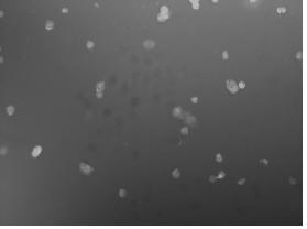
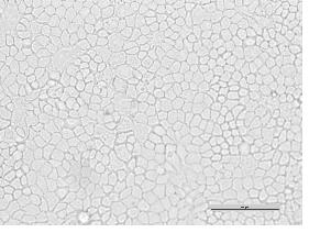
Method for marking canine kidney cells through chitosan-quantum dot fluorescent probe
A technology of dog kidney cells and fluorescent probes, applied in fluorescence/phosphorescence, material excitation analysis, etc., to achieve the effects of easy operation, controllable fluorescence intensity and color, and good biocompatibility
- Summary
- Abstract
- Description
- Claims
- Application Information
AI Technical Summary
Problems solved by technology
Method used
Image
Examples
Embodiment 1
[0032] A method for labeling dog kidney cells with a chitosan-quantum dot fluorescent probe, comprising the following steps:
[0033] 1. Prepare 0.0005mol / L green and red carboxymethyl chitosan-CdTe quantum dot solutions:
[0034] Pipette 2.5mL of 0.002mol / L green and red carboxymethyl chitosan-CdTe quantum dot solutions into sterilized 10mL volumetric flasks, add high-purity water (resistivity greater than 18 MΩ·cm) to volume ,spare. The selected chitosan-quantum dot fluorescent probe is formed by self-assembly of carboxymethyl chitosan and CdTe quantum dots in the aqueous phase through electrostatic complexation, covalent binding, chelation, etc., and has good water solubility. and biocompatibility; (carboxymethyl chitosan-CdTe quantum dots are self-made, and the brief preparation process is as follows: CdCl 2 The solution and the thioglycolic acid solution were mixed evenly, and the pH value was adjusted with NaOH solution to be alkaline, and then newly prepared KTeH was ...
Embodiment 2
[0042] A method for labeling dog kidney cells with a chitosan-quantum dot fluorescent probe, the steps are as follows:
[0043] 1. Prepare 0.0001mol / L carboxymethyl chitosan-CdTe quantum dot solution:
[0044] Pipette 0.5mL of 0.002mol / L yellow-green carboxymethyl chitosan-CdTe quantum dot solution into a sterilized 10mL volumetric flask, add high-purity water (resistivity greater than 18 MΩ·cm) to volume, and set aside . The selected chitosan-quantum dot fluorescent probe is formed by self-assembly of carboxymethyl chitosan and CdTe quantum dots in the aqueous phase through electrostatic complexation, covalent binding, chelation, etc., and has good water solubility. and biocompatibility;
[0045] 2. Culture of dog kidney cells:
[0046] The dog kidney cells (MDCK) that were full and in good growth condition in the T25 cell culture flask were digested with 0.05% EDTA-trypsin into a single cell suspension, and 1 × 10 cells were taken after counting the cells. 5 The dog kidn...
Embodiment 3
[0053] A method for labeling dog kidney cells with a chitosan-quantum dot fluorescent probe, the steps are as follows:
[0054] 1. Prepare 0, 0.00005, 0.0001, 0.00025, 0.0005, 0.001mol / L carboxymethyl chitosan-CdTe quantum dot solution:
[0055] Pipette 0, 0.25, 0.5, 1.25, 2.5, 5mL of 0.002mol / L yellow-green carboxymethyl chitosan-CdTe quantum dot solution into a sterilized 10mL volumetric flask, add high-purity water (resistivity greater than 18 MΩ·cm) constant volume, spare. The selected chitosan-quantum dot fluorescent probe is formed by self-assembly of carboxymethyl chitosan and CdTe quantum dots in the aqueous phase through electrostatic complexation, covalent binding, chelation, etc., and has good water solubility. and biocompatibility;
[0056] 2. Culture of dog kidney cells:
[0057] The dog kidney cells (MDCK) that were full and in good growth condition in the T25 cell culture flask were digested with 0.05% EDTA-trypsin into a single cell suspension, and 1 × 10 ce...
PUM
| Property | Measurement | Unit |
|---|---|---|
| electrical resistivity | aaaaa | aaaaa |
| fluorescence | aaaaa | aaaaa |
Abstract
Description
Claims
Application Information
 - R&D
- Intellectual Property
- Life Sciences
- Materials
- Tech Scout
- Unparalleled Data Quality
- Higher Quality Content
- 60% Fewer Hallucinations
Browse by: Latest US Patents, China's latest patents, Technical Efficacy Thesaurus, Application Domain, Technology Topic, Popular Technical Reports.
© 2025 PatSnap. All rights reserved.Legal|Privacy policy|Modern Slavery Act Transparency Statement|Sitemap|About US| Contact US: help@patsnap.com


