
Fluorescence spectrometry method for oncogene C-myc protein

An oncogene and protein technology, applied in the field of protein content detection in tumor cells, can solve the problems of application limitation, cumbersome operation, low precision, etc., and achieve the effect of fast universality and simple operation.

Inactive Publication Date: 2010-11-24
CHANGSHA UNIVERSITY OF SCIENCE AND TECHNOLOGY
View PDF1 Cites 2 Cited by
  • Summary
  • Abstract
  • Description
  • Claims
  • Application Information

AI Technical Summary

Problems solved by technology

These methods are cumbersome to operate and have low precision, which limits their application. Therefore, it is necessary to find a method that can quickly and quantitatively detect C-myc protein

Method used


Image

  • Fluorescence spectrometry method for oncogene C-myc protein
  • Fluorescence spectrometry method for oncogene C-myc protein
  • Fluorescence spectrometry method for oncogene C-myc protein

Examples


Embodiment 1

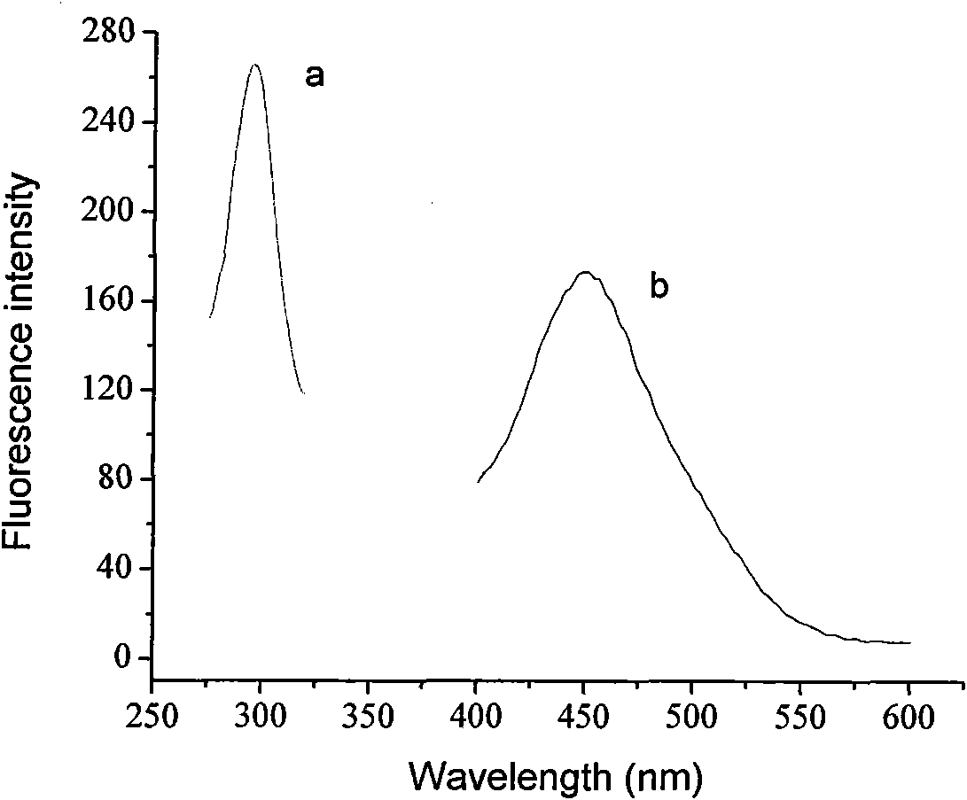
[0021] Select the scanning conditions for the C-myc standard sample, and determine the excitation wavelength and emission wavelength.

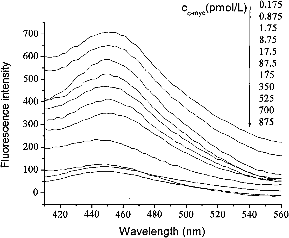
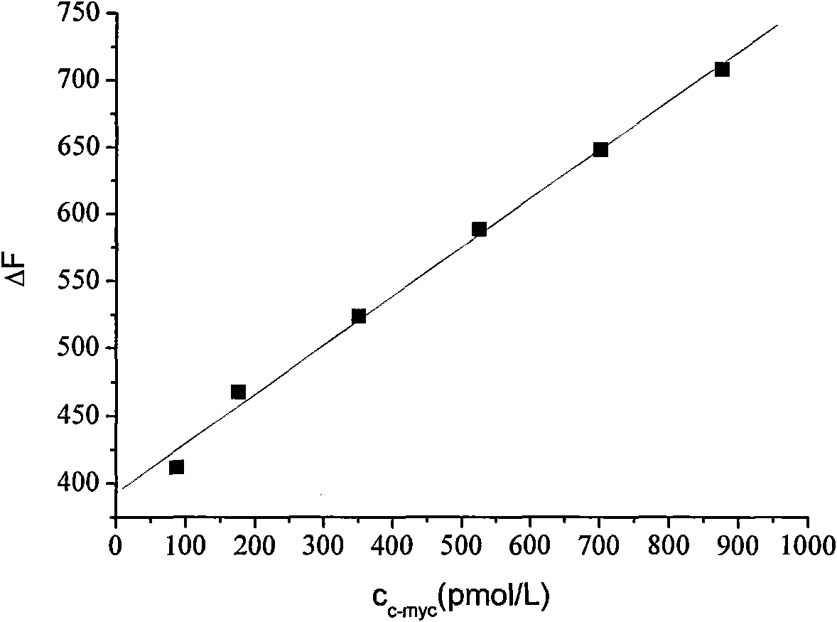
[0022] Using PBS buffer as a solvent and 1 μL of gold sol with a certain particle size (12nm, 64pmol / L) as a matrix, a series of concentration C-myc standard solutions were prepared. Set the scan range to 200-600nm. According to the concentration from low to high, add a certain amount of standard solution to the cuvette for measurement, take the C-myc concentration as the abscissa, and the fluorescence quenching intensity ΔF as the ordinate, draw the standard curve for measuring the concentration of the C-myc sample (see image 3 and 4 ).

Embodiment 2

[0024] In a 1mL cuvette, prepare the Au-C-myc-PBS mixture (C c-myc =437.5pmol / L, C Au =128pmol / L, Φ Au =21nm), set the scan range to 300-500nm, under the condition of 4°C, fix the excitation wavelength, measure the fluorescence emission intensity every 5 minutes, measure for 30 minutes, the results show the relative standard deviation of the fluorescence intensity obtained under the set experimental conditions It is 0.38%, indicating that the stability of this method is good.

Embodiment 3

[0026] In a 1mL cuvette, prepare the Au-C-myc-PBS mixture (Φ Au =32nm,C Au =256pmol / L), set the scanning range 300-500nm, fix the excitation wavelength, and repeatedly measure the fluorescence intensity of two different concentrations of C-myc samples (87.5pmol / L and 437.5pmol / L) for 12 times (the interval is 2 minutes) , the results show that the relative standard deviations are only 0.50% and 0.87% under the set experimental conditions, which shows that the method has good reproducibility and ensures the accuracy of the measured experimental data.


PUM


Abstract

The invention discloses a fluorescence spectrometry method for an oncogene C-myc protein. In the method, the determination basis is that the C-myc protein has fluorescence and has a fluorescence quenching phenomenon after combining with gold nano-sol; and the fluorescence quenching intensity (delta F) and the concentration change of the C-myc protein are in a linear corresponding relationship so as to fulfill the aim of determining the C-myc content. A C-myc protein sample is determined accurately by the method, the recovery rate of the C-myc is between 94.29 and 107.14 percent; the detectable C-myc protein concentration range is between 0 and 1,000 pmol / L; the linear range is between 87.5 and 875 pmol / L and between 0.175 and 17.5 pmol / L; and the detection limit is between 0.01 and 0.08 pmol / L. The determination method of the invention has the advantages of simple and convenient operation, wide linear range, high sensitivity, low detection limit, high stability and reproducibility and the like.

Description

technical field [0001] The invention belongs to the technical field of protein content detection in tumor cells, and in particular relates to a fluorescence spectrum method for detecting oncogene C-myc recombinant protein. Background technique [0002] C-myc protein is one of the earliest proto-oncogene products found to be related to the proliferation activity of tumor cells. It is located in the nucleus and plays an important role in regulating DNA synthesis, cell proliferation, apoptosis, differentiation and cell cycle progression. Important role, but also closely involved in the transformation of cell tumors. The expression of C-myc protein is closely related to the initiation and cancerous degree of many cancerous tissues and cells. [0003] At present, there are traditional biological enzyme-linked immunosorbent assays (Enzyme-linked immunosorbent assay, ELISA) for the detection of C-myc protein, and traditional methods can only be qualitative or semi-quantitative. T...

Claims


Application Information

Patent Timeline
IPC IPC(8): G01N21/64G01N33/53
Inventor 曹忠吕品龙姝王明星戴云林马贵福谭淑珍
Owner CHANGSHA UNIVERSITY OF SCIENCE AND TECHNOLOGY
Learn More