Artificial nasolacrimal canal
A nasolacrimal duct and artificial technology, applied in the medical field, can solve problems such as inability to treat lacrimal duct blockage, increase in surgical failure rate, and inability to use guided pull-in surgery, so as to improve safety factor, service life, and indications. Wide, curative effect
- Summary
- Abstract
- Description
- Claims
- Application Information
AI Technical Summary
Problems solved by technology
Method used
Image
Examples
Embodiment 1
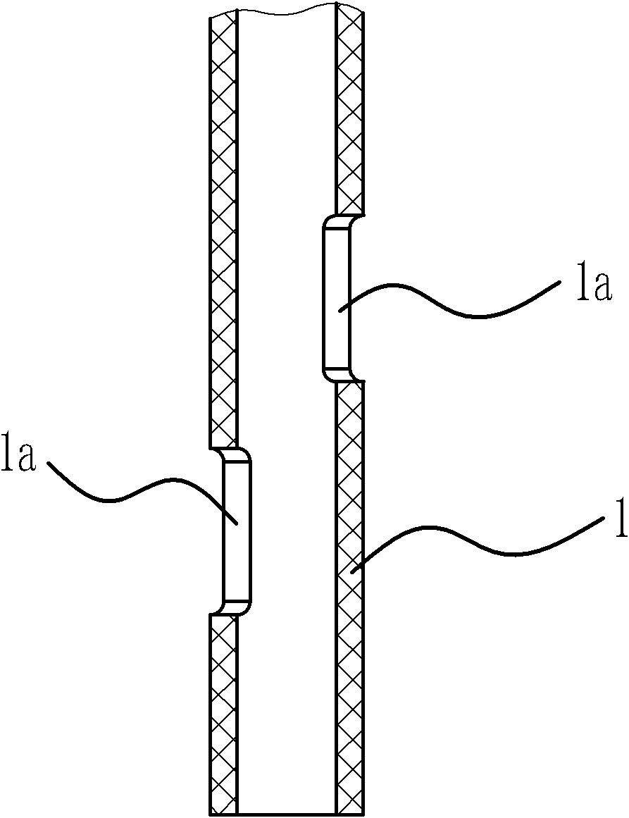
[0036] Such as Figure 1 to Figure 4 As shown, the artificial nasolacrimal duct includes a drainage main pipe 1 , a fixed flap 2 , a fixed cap 3 , and a drainage branch pipe 4 .
[0037] Specifically, as figure 1 with figure 2As shown, the drainage main pipe 1 is in the shape of a circular tube, and a positioning hole 1a is opened in its lower part. The position of the positioning hole 1a relative to the drainage branch tube 4 is determined, so only need to determine the positional relationship of the positioning hole 1a relative to the nasal passage during the operation, that is, the drainage branch tube 4 can be accurately introduced into the lacrimal canaliculus. If the artificial nasolacrimal duct fails to reach the designated position after introduction, the artificial nasolacrimal duct can be pulled out with a guide wire hook at the positioning hole 1a. Therefore, setting the positioning hole 1a can make the operation more convenient, safe and accurate.
[0038] The...
Embodiment 2
[0046] Such as Figure 5 with Figure 7 As shown, the structure and principle of this embodiment are basically the same as that of Embodiment 1, the difference is that according to the physiological structure of the nasolacrimal duct of the human body, when the fixed valve 2 is located in the lacrimal sac, the extension line of the upper lacrimal canaliculus is located at the fixed position. Petal 2 and close to the fixed cap 3. Therefore, in order to enable the above-mentioned drainage branch tube 4 to penetrate into the upper lacrimal canaliculus, while the drainage branch tube 4 does not deform the upper lacrimal canaliculus and the position of the drainage branch tube 4 relative to the fixed cap 3 does not change, the drainage branch tube 4 The rear end is fixedly connected on the lower end surface of the fixed cap 3. The junction of the drainage branch tube 4 and the fixed cap 3 is located at one of the gaps between the two fixed flaps 2, but only occupies a small area,...
Embodiment 3
[0050] Such as Image 6 with Figure 8 As shown, the structure and principle of this embodiment are basically the same as that of Embodiment 1, the difference is that: at one of the gaps between the two fixed flaps 2, there are two tubular drainage branches that can penetrate into the lacrimal canaliculus 4, that is, the first drainage branch pipe 41 and the second drainage branch pipe 42.
[0051] The lacrimal canaliculus includes the upper canaliculus and the small canaliculus; according to the physiological structure of the human nasolacrimal duct, when the fixed flap 2 is located in the lacrimal sac, the extension of the lower canaliculus is located at the fixed flap 2 and close to the drainage tube 1, and the upper lacrimal canaliculus The extension of the tubule is located at and close to the fixed flap 2. So in order to make the above-mentioned drainage branch tube 1 41 penetrate into the lower lacrimal canaliculus, the drainage branch tube 2 42 can penetrate into the...
PUM
 Abstract
Description
Claims
Application Information
 - R&D
- Intellectual Property
- Life Sciences
- Materials
- Tech Scout
- Unparalleled Data Quality
- Higher Quality Content
- 60% Fewer Hallucinations
Browse by: Latest US Patents, China's latest patents, Technical Efficacy Thesaurus, Application Domain, Technology Topic, Popular Technical Reports.
© 2025 PatSnap. All rights reserved.Legal|Privacy policy|Modern Slavery Act Transparency Statement|Sitemap|About US| Contact US: help@patsnap.com


