II type collagen sponge bracket and uses thereof
A collagen sponge and collagen technology, which is applied in the field of biomedicine, can solve the problems of difficulty in material scaffold forming research and animal experiments, difficult to use as a cell culture scaffold material, and complicated extraction method of type II collagen, etc., achieves good repair effect, and increases three-dimensional stability. The effect of improving the elasticity and elastic modulus
- Summary
- Abstract
- Description
- Claims
- Application Information
AI Technical Summary
Problems solved by technology
Method used
Image
Examples
Embodiment 1
[0031] 1. Preparation of high-purity type Ⅱ collagen [Reference: Li Siming, Ye Chunting, etc., Preparation and detection of high-purity porcine cartilage type Ⅱ collagen, Journal of Biomedical Engineering, 2001, 18 (4) 592-594]:
[0032] Material:
[0033] Fresh pig articular cartilage: obtained from the limb articular cartilage of freshly slaughtered pigs in the slaughterhouse
[0034] Guanidine hydrochloride: Farco 500g / bottle
[0035] Pepsin: Sigma 100g / bottle
[0036] The rest are domestic analytical reagents.
[0037] method:
[0038] Cut the hyaline cartilage from freshly slaughtered pig limb joints, cut into thin slices, degrease, mash, and homogenate, then stir with 10 times the volume of 4M (pH7.5) guanidine hydrochloride for 24 hours, and centrifuge; the sediment diameter is fully washed , Weigh the precipitated cartilage particles with a wet weight of about 260g, hydrolyze with pepsin at a mass ratio of 1 / 50 under acidic conditions for 24 to 48 hours, centrifuge...
experiment example 1
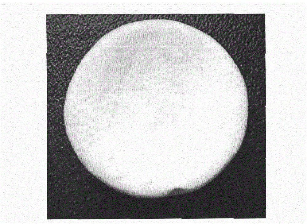
[0044] Experimental example 1. Pore diameter detection of type II collagen sponge scaffold material
[0045] The type II collagen sponge material was prepared according to the above method, and divided into stock solution non-crosslinked group, stock solution crosslinked group, concentrated non-crosslinked group, and concentrated crosslinked group.
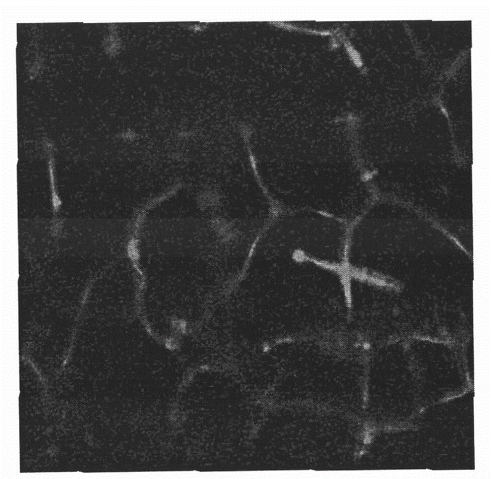
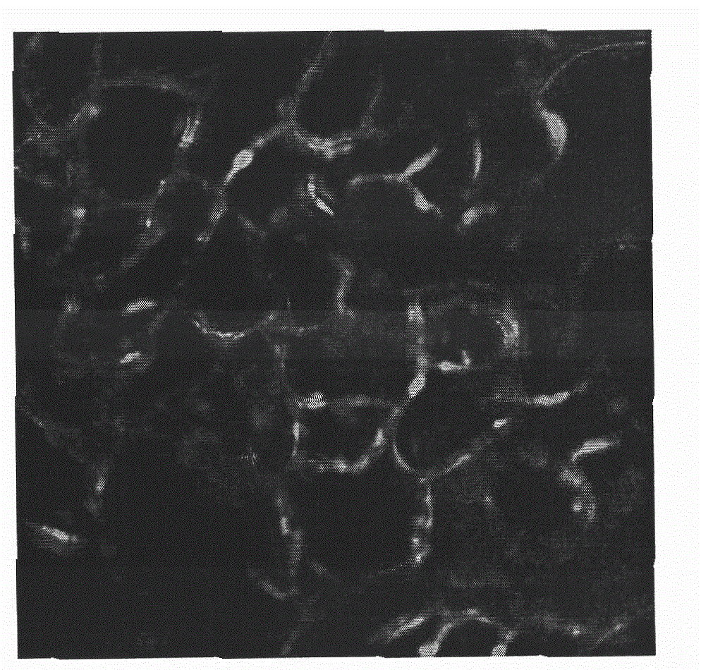
[0046] Collagen autofluorescence layered scanning of collagen sponges with various pore diameters was carried out by laser confocal microscope. The excitation wavelength was 488nm, and the emission wavelength was 520nm. The autofluorescence signal intensity was measured to determine the pore size of the sponge material; using CLSM image The analysis system is used for operation and analysis, and then the SPSS13.0 software package is used to make statistics on the data.
[0047] As shown in Figure 2, the laser confocal microscopy results show that the collagen sponges of each group have obvious autofluorescence, indicating that the...
experiment example 2
[0051] Experimental Example 2: Solubility Detection of Type II Collagen Scaffold Materials
[0052] The type II collagen sponge material was prepared according to the above method, which was divided into a cross-linked group and an uncross-linked group.
[0053] Cut the type II collagen sponge material into 1×1cm 2 Put it into a 6-well culture plate, add 3ml of DMEM medium, and observe the condition of the sponge at 37°C.
[0054] The results showed that the non-cross-linked type II collagen sponge material swelled after being added to the culture medium, and partially dissolved, and the material became soft and translucent; after 7 days, the material was completely dissolved. The appearance of the cross-linked sponge material remains unchanged after being added to the culture medium, and it can still maintain a complete state up to 50 days after being planted into the cells.
PUM
| Property | Measurement | Unit |
|---|---|---|
| pore size | aaaaa | aaaaa |
| pore size | aaaaa | aaaaa |
| pore size | aaaaa | aaaaa |
Abstract
Description
Claims
Application Information
 - R&D
- Intellectual Property
- Life Sciences
- Materials
- Tech Scout
- Unparalleled Data Quality
- Higher Quality Content
- 60% Fewer Hallucinations
Browse by: Latest US Patents, China's latest patents, Technical Efficacy Thesaurus, Application Domain, Technology Topic, Popular Technical Reports.
© 2025 PatSnap. All rights reserved.Legal|Privacy policy|Modern Slavery Act Transparency Statement|Sitemap|About US| Contact US: help@patsnap.com


