Therapeutic agent for acceleration of spinal nerve repair comprising ghrelin, derivative thereof or substance capable of acting on GHS-R1a as active ingredient
A technology of spinal nerves and therapeutic agents, applied in the field of proliferation of spinal nerve precursor cells
- Summary
- Abstract
- Description
- Claims
- Application Information
AI Technical Summary
Problems solved by technology
Method used
Image
Examples
Embodiment 1
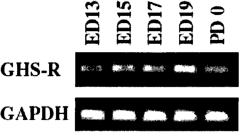
[0230] Example 1 Expression of GHS-R1a mRNA in Rat Fetal Spinal Cord
[0231] Spinal cord tissues were excised from Wistar rat fetuses on days 13 to 19 of pregnancy and rat fetuses just after birth, using Trizol reagent (Life Technologies, Inc., Gaithersburg, MD USA), according to Nakahara et al.: Biochem.Biophys.Res. Total RNA was extracted by the method described in Commun. 303: 751-755 (2003). 1 μg of total RNA was reverse-transcribed with random primers, and single-stranded cDNA was synthesized using Superscript 3 preamplification reagent (Life Technologies, Inc.). Using the sense and antisense primers specific for GHS-R1a, the PCR method (using BDAdvantage TM 2 PCR Enzyme System, BD Science, CA USA) After amplifying the obtained cDNA, electrophoresis was performed using 2% agarose gel. In addition, as a control mRNA, GAPDH (glyceraldehyde-3-phosphate dehydrogenase) stably expressed in cells was used.
[0232] As PCR primers specific to GHS-R1a, 5'-GATACCTCTTTTTCCAAG...
Embodiment 2
[0235] Example 2 Existence of GHS-R1a in spinal cord cells
[0236] Fetal spinal cords were collected from 17-day pregnant Wistar rats, and frozen sections with a thickness of 14 μm were prepared. The sections were fixed with 4% paraformaldehyde / 0.1 M phosphate buffer for 30 minutes, washed with 0.1 M phosphate buffer, and incubated with 2% normal goat serum in PBS for 30 minutes at room temperature. Then, the sections were washed 3 times with PBS, incubated overnight at 4°C with rabbit anti-GHS-R antibody, washed with PBS, incubated with goat anti-rabbit IgG conjugated to Alexa Fluoro 488, and immunostained. After washing the residual antibody, the sections were embedded and observed with a fluorescence microscope.
[0237] The result is as figure 2 shown. According to immunostaining using an anti-GHS-R antibody, it was shown that in the gray matter where the cell bodies of neurons are present, GHS-R1a ( figure 2 A). After pretreatment with anti-GHS-R antibody, the i...
Embodiment 3
[0238] During the cell proliferation of embodiment 3, nestin in spinal cord nerve cells and spinal cord neural precursor cells, Coexistence of Map2 and GHS-R 1a
[0239] Double immunostaining was performed to confirm the coexistence of Nestin, a marker of neuron precursor cells, and Map2, and GHS-R, a marker of neuron cells, in proliferating cells (cells incorporating BrdU).
[0240] Fetus were removed by laparotomy under anesthesia from gestational 17-day Wistar rats. The spinal cord was collected from the fetus, digested with papain in cold Hanks solution, and mechanically separated by a pipette to obtain a fetal spinal cord cell dispersion. After the dispersed cells were filtered and centrifuged, they were suspended in NaHCO-containing 3 , 5% fetal calf serum, penicillin (100 U / mL) and streptomycin (100 μg / mL) in DMEM medium, on a laminin-coated 96-well multi-well plate in 10 5 Cells / well Seed cells.
[0241] 5-Bromo-2'-deoxyuridine (BrdU) (10 µM) was added thereto a...
PUM
 Abstract
Description
Claims
Application Information
 - Generate Ideas
- Intellectual Property
- Life Sciences
- Materials
- Tech Scout
- Unparalleled Data Quality
- Higher Quality Content
- 60% Fewer Hallucinations
Browse by: Latest US Patents, China's latest patents, Technical Efficacy Thesaurus, Application Domain, Technology Topic, Popular Technical Reports.
© 2025 PatSnap. All rights reserved.Legal|Privacy policy|Modern Slavery Act Transparency Statement|Sitemap|About US| Contact US: help@patsnap.com


