
Imaging apparatus, medical image processing apparatus, and medical image processing method

A camera device and medical image technology, applied in the field of display of tumor nutrient blood flow, can solve the problem of no provision

Active Publication Date: 2009-08-26
TOSHIBA MEDICAL SYST CORP
View PDF2 Cites 8 Cited by
  • Summary
  • Abstract
  • Description
  • Claims
  • Application Information

AI Technical Summary

Problems solved by technology

However, no means are provided in the prior art to efficiently display such images

Method used


Image

  • Imaging apparatus, medical image processing apparatus, and medical image processing method
  • Imaging apparatus, medical image processing apparatus, and medical image processing method
  • Imaging apparatus, medical image processing apparatus, and medical image processing method

Examples


Embodiment Construction

[0020] Hereinafter, preferred embodiments of the medical imaging device, ultrasonic imaging device, magnetic resonance imaging device, medical image processing device, and medical image processing method of the present invention will be described in detail with reference to the accompanying drawings.

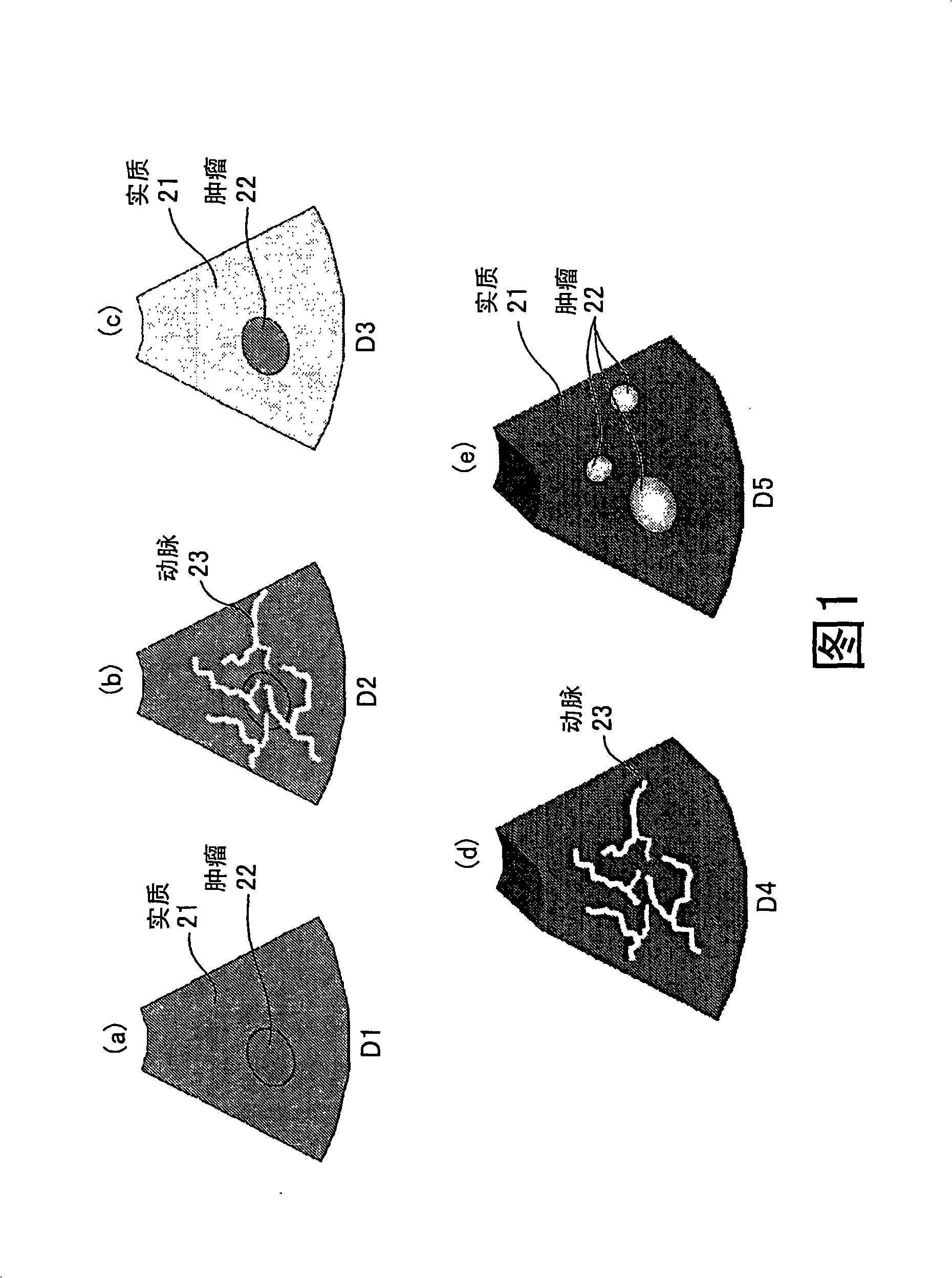
[0021] First, a synthesized image displayed by the ultrasonic diagnostic apparatus according to the present embodiment will be described. The ultrasound diagnostic apparatus of the present embodiment collects three pieces of data with different phases in contrast-enhanced ultrasound, and generates and displays a composite image. figure 1 It is an explanatory diagram for explaining a synthesized image displayed by the ultrasonic diagnostic apparatus of this embodiment.

[0022] figure 1 (a) represents the B-mode image 'D1' before or immediately after contrast agent injection, figure 1 (b) is the B-mode image 'D2' in the arterial phase after contrast injection, figure 1 (c) sho...


PUM


Abstract

The invention provides a medial imaging apparatus, ultrasonic imaging apparatus, magnetic resonance imaging apparatus, medical image processing apparatus and medical image processing method. The differential-image creating unit (7) creates a three-dimensional image D4 on which an artery (23) is highlighted and a three-dimensional image D5 on which a tumor (22) is highlighted. An image compositing unit (8) combines the three-dimensional images D4, D5 to composite an image. A display control unit (9) displays the composite image on a monitor (11). Moreover, when compositing an image, the image compositing unit (8) composites the image such that a tumor nutrient blood-flow (24) inside and outside tumor is to be displayed in respective different colors. Furthermore, a blood-flow quantity inside tumor measuring unit calculates a blood-flow quantity inside tumor and a ratio of the blood-flow quantity inside tumor to a tumor volume, and the display control unit (9) displays the blood-flow quantity inside tumor and the ratio of the blood-flow quantity inside tumor to the tumor volume. Accordingly, information that is meaningful for determining malignancy of a tumor can be provided.

Description

[0001] The present invention enjoys the right of priority of Japanese Patent Application (Application No. 2008-024296) filed on February 4, 2008, and the entire content of the Japanese Patent Application is cited in this application. technical field [0002] The present invention relates to a technique for displaying tumor nutrient blood flow in tumor imaging using a contrast medium. Background technique [0003] In recent years, ultrasound contrast agents based on microbubbles that enhance echo reflection in intravenous injection have been commercialized, and contrast echo methods have been implemented. According to this method, blood flow dynamics can be clearly observed by enhancing blood flow signals with microbubbles introduced into blood vessels. Furthermore, microvesicles have a specific property of being phagocytosed by Kupffer cells in the liver, and are expected to be used in the field of liver tumor diagnosis. [0004] In the contrast echo method, the image to be...

Claims


Application Information

Patent Timeline
Patent Type & Authority Applications(China)
IPC IPC(8): A61B8/14A61B8/06A61B5/055
CPCA61B8/13A61B8/08A61B8/463A61B8/481A61B8/488A61B8/06A61B8/5238
Inventor 青柳康太山形仁江马武博佐藤恭子佐藤俊介
Owner TOSHIBA MEDICAL SYST CORP
Learn More