Accurate positioning method based on atomic force microscope

An atomic force microscope and precise positioning technology, which is applied to the parts of the instrument, instruments, surface/boundary effects, etc., can solve the problems of time-consuming consumables, insufficient positioning, and substrate damage scales, etc., to avoid time-consuming consumables, The effect of simple, fast and precise positioning

Inactive Publication Date: 2009-04-22
WUHAN UNIV
View PDF2 Cites 7 Cited by
  • Summary
  • Abstract
  • Description
  • Claims
  • Application Information

AI Technical Summary

Problems solved by technology

For an atomic force microscope without a supporting optical system, it is difficult to precisely locate a specific position
The Chinese patent "Scanning Probe Microscope Sample Positioning Method" of Patent No. 99126338.3 reports a method, which is to divide a small area on one side of a transparent substrate and mark different numbers, foreign letters or various digital symbols and other identifiers to show difference, or after marking various identifiers in the small squares of the coordinate axis graph paper, paste them on the reverse side of the transparent substrate with double-sided tape or glue, or use etching technology to etch the coordinates on one side of the transparent substra

Method used


Image

  • Accurate positioning method based on atomic force microscope
  • Accurate positioning method based on atomic force microscope
  • Accurate positioning method based on atomic force microscope

Examples


Embodiment 1

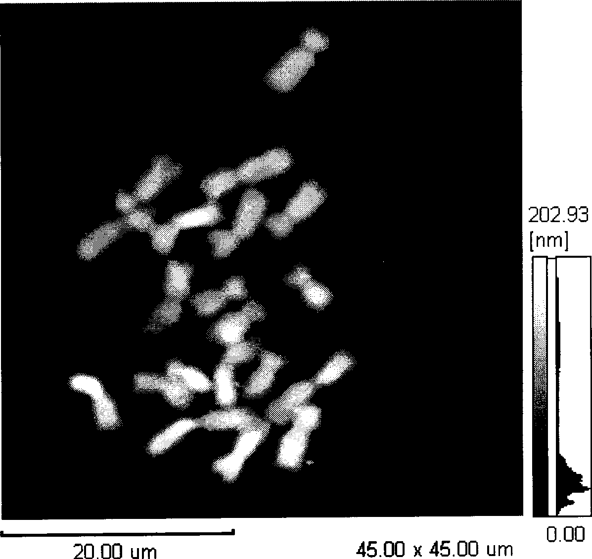
[0019] Observation of maize metaphase chromosomes by atomic force microscope.

[0020] 1. Conventional chromosome preparation method Maize metaphase chromosomes were prepared on glass slides.

[0021] 2. Use an optical phase contrast microscope to find the chromosome to be tested.

[0022] 3. Under the optical microscope, the copper sheet PELCO Position the Center-Marked Grids above the sample to be tested, record the area where the sample is located, and use the adsorption force to place the PELCO Center-Marked Grids are sucked onto the substrate of the sample.

[0023] 4. Attach the PELCO The base of Center-Marked Grids is attached to the stage of atomic force microscope with double-sided tape.

[0024] 5. Scanning PELCO with AFM Center-Marked Grids, the scanning range is reduced step by step, and the probe of the atomic force microscope is positioned above the corresponding grid of the sample to be tested.

[0025] 6. Use the ear wash ball to put the PELCO Cente...

Embodiment 2

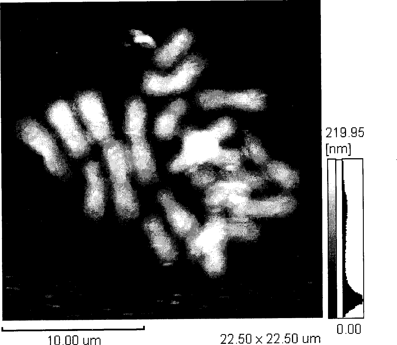
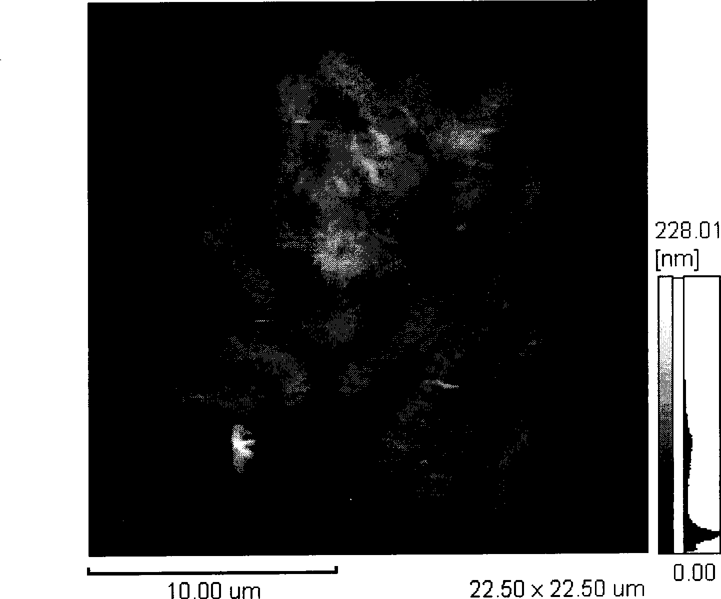
[0027] Comparison of Maize Metaphase Chromosomes Before and After DNase I Treatment Using Atomic Force Microscopy

[0028] 1. Conventional chromosome preparation method Maize metaphase chromosomes were prepared on glass slides.

[0029] 2. Use an optical phase contrast microscope to find the chromosome to be tested.

[0030] 3. Under the optical microscope, the copper mesh PELCO Position the Center-Marked Grids above the sample to be tested, record the area where the sample is located, and use the adsorption force to place the PELCO Center-Marked Grids are sucked onto the substrate of the sample.

[0031] 4. Attach the PELCO The base of Center-Marked Grids is attached to the stage of atomic force microscope with double-sided tape.

[0032] 5. Scanning PELCO with AFM Center-Marked Grids, the scanning range is reduced step by step, and the probe of the atomic force microscope is positioned above the corresponding grid of the sample to be tested.

[0033] 6. Use the ear...


PUM


Abstract

The invention discloses a method for precise orientation by an atomic force microscope. The precise orientation method comprises steps of: firstly finding a sample to be measured by an optical microscope; putting a copper net Center_Marked Grids above the sample to be measured, the copper net being formed by ruled grids with the same size, recording the region of the grids corresponding to the sample to be measured under the optical microscope, scanning the Center_Marked Grids by using an atomic force microscope, reducing the scanning area stage by stage, locating a probe of the atomic force microscope above the corresponding grids of the sample to be measured; and finally, softly blowing the Center_Marked Grids from side by using a washing ear ball; thereby carrying out the precise observation of the atomic force microscope by knitting stitch. The method can precisely orientate the sample to be scanned.

Description

technical field [0001] The invention relates to an accurate positioning method based on an atomic force microscope, that is, a method for accurately positioning a sample in a micro region by using an atomic force microscope. technical background [0002] AFM is an important tool for studying the surface structure of objects at the nanometer scale, and has been widely used in physics, biology, chemistry and other research. At the beginning of the invention of the atomic force microscope, it has been used for physical research. The universal uniformity of physical materials determines that it is not necessary to image a specific sample, so that it is not necessary to accurately position the sample to be tested. Therefore, the atomic force microscope of some manufacturers does not have a supporting optical system ( Such as the 9500J3 atomic force microscope of Shimadzu Corporation of Japan, and the Dimension 3100 atomic force microscope of Veeco Precision Instrument Co., Ltd. o...

Claims


Application Information

Patent Timeline
IPC IPC(8): G01N13/16G12B21/20G01Q10/00
CPCG01Q40/00
Inventor 李立家马璐
Owner WUHAN UNIV
Why Eureka
Try Eureka