
Method for Measuring Cell Cycle Using Cell Autofluorescence Spectroscopy

A technology of cell cycle and autofluorescence, which is applied in the direction of fluorescence/phosphorescence, biochemical equipment and methods, measurement/inspection of microorganisms, etc., to achieve the effects of eliminating side effects, convenient operation, and simple sample preparation

Inactive Publication Date: 2012-02-01
CHONGQING UNIV
View PDF0 Cites 0 Cited by
  • Summary
  • Abstract
  • Description
  • Claims
  • Application Information

AI Technical Summary

Problems solved by technology

[0005] At present, there are no reports at home and abroad to judge the cell cycle of cells by establishing a cell autofluorescence spectrum model

Method used


Image

  • Method for Measuring Cell Cycle Using Cell Autofluorescence Spectroscopy
  • Method for Measuring Cell Cycle Using Cell Autofluorescence Spectroscopy
  • Method for Measuring Cell Cycle Using Cell Autofluorescence Spectroscopy

Examples


Embodiment

[0016] Example: Method for cell cycle measurement of Hela cells using fluorescence spectroscopy:

[0017] 1. First, establish the fluorescence spectrum model of the Hela cell cycle, and obtain the cell standard spectrum curve of each phase according to the following steps:

[0018] ① Acquisition of samples. In order to obtain accurate spectral data, it is first necessary to ensure that the cell sample is in a natural growth state, and use an optical microscope to observe the morphological changes to obtain single cells in different phases of the cell cycle (G1, S, G2, and M). Use it as a standard sample.

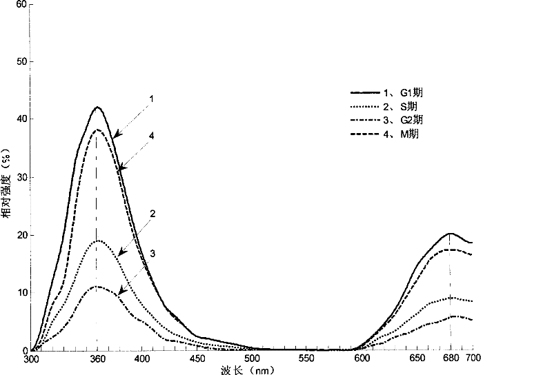
[0019] ②Acquisition of the best excitation wavelength of cells in each phase. First scan with 250nm as the excitation wavelength in the range of 250~700nm to obtain the emission spectrum of the cell at 250nm. After measuring the excitation spectrum from 250nm to the first fluorescence peak wavelength in the emission spectrum at 360nm, find the fluorescence in the excitation spect...


PUM


Abstract

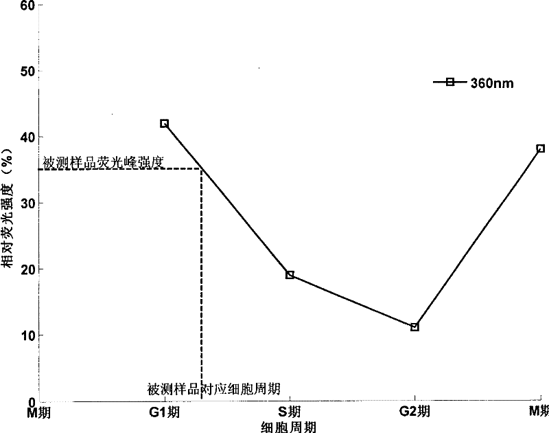
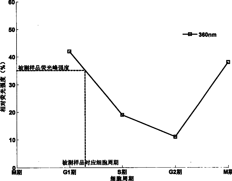
The invention discloses a method for measuring cell cycle by using cell autofluorescence spectrum. This method utilizes the peak position, spectral peak, and Based on the influence of the peak intensity, find out the corresponding relationship between these parameters and the cell cycle, establish the cell cycle fluorescence spectrum model of a specific cell, and finally use this model to achieve accurate determination of a single cell cycle. The present invention has simple sample preparation, convenient operation, common equipment, high accuracy, and fast measurement speed. By applying the relationship between cell absorption peak intensity and cell cycle variation, the cell cycle of cells can be judged conveniently, and cell samples do not need to use fluorescent light. The dye pair stains, thereby eliminating the side effects caused by the use of fluorescent dyes, so that the cells can be used for subsequent culture and research after measurement.

Description

Technical field [0001] The invention relates to a method for measuring cell cycle, in particular to a method for accurately reflecting cell cycle changes using cell autofluorescence spectra. Background technique [0002] Cells are the basis of life science research, and all key issues of life must be found in cells. The research of biological reproductive development, heredity, nerve (brain) activity and other major life phenomena must be based on cells. The growth and development of plants and animals is achieved by cell proliferation, cell differentiation and cell apoptosis. The pathogenesis of all diseases should also be based on cytopathic research. Cell division is the most basic and important process of the origin of life. Cell growth and cell division are the basic forms of all biological proliferation, and the cycle of cell growth and cell division is the cell proliferation cycle referred to as the cell cycle. For eukaryotic cells, the cell cycle consists of G1 phase, ...

Claims


Application Information

Patent Timeline
Patent Type & Authority Patents(China)
IPC IPC(8): G01N33/48G01N21/64C12Q1/02
Inventor 林晓钢潘英俊郭永彩高潮
Owner CHONGQING UNIV
Learn More