Three-dimensional modeling method for funnel chest of children
A three-dimensional modeling, pectus excavatum technology, applied in 3D modeling, image data processing, instruments, etc., can solve the problems of poor spiral CT image quality, invisible cartilage, invisible cartilage CT images, etc., to achieve a high degree of simulation. Effect
- Summary
- Abstract
- Description
- Claims
- Application Information
AI Technical Summary
Problems solved by technology
Method used
Image
Examples
Embodiment Construction
[0023] The establishment method of pectus excavatum thorax three-dimensional model of the present invention, specifically carries out according to the following steps:
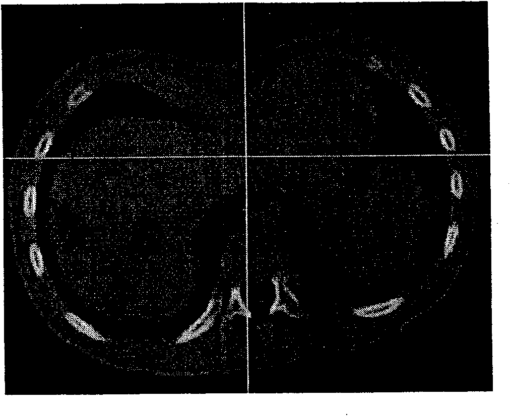
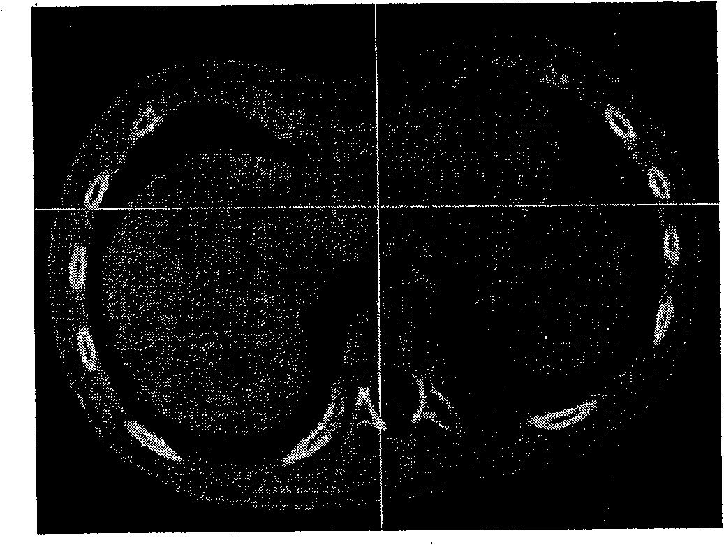
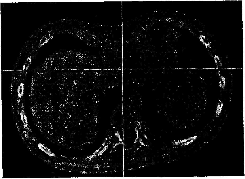
[0024] Will figure 1 The original spiral CT image of pectus pectus excavatum is input into the computer. CT images are sliced images, displayed in grayscale mode, divided into three directions: front view, top view, and left view. The image resolution is 512×512 pixels, the number of slices is 227, and the interval between slices is 0.4 mm. From the gray scale of the original CT image, only the difference between rib hard bone and soft tissue can be distinguished, but cartilage and soft tissue cannot be distinguished.
[0025] Firstly, image processing was performed on the original spiral CT images of pectus pectus excavatum in a child. For the case that the original spiral CT image of pectus pectus excavatum is a grayscale image, the costal cartilage grayscale data was extracted first. The gray value ra...
PUM
 Abstract
Description
Claims
Application Information
 - R&D
- Intellectual Property
- Life Sciences
- Materials
- Tech Scout
- Unparalleled Data Quality
- Higher Quality Content
- 60% Fewer Hallucinations
Browse by: Latest US Patents, China's latest patents, Technical Efficacy Thesaurus, Application Domain, Technology Topic, Popular Technical Reports.
© 2025 PatSnap. All rights reserved.Legal|Privacy policy|Modern Slavery Act Transparency Statement|Sitemap|About US| Contact US: help@patsnap.com


