Accelerated magnetic resonance thermometry

a magnetic resonance thermometer and accelerated technology, applied in the field of magnetic resonance imaging, can solve the problems of not giving absolute temperature, method is too slow to be used in continuous imaging for most applications,

Active Publication Date: 2018-05-15
KONINKLIJKE PHILIPS ELECTRONICS NV
View PDF27 Cites 4 Cited by
  • Summary
  • Abstract
  • Description
  • Claims
  • Application Information

AI Technical Summary

Benefits of technology

[0011]Both methods, the PRF and PS method suffer from different weaknesses. Combining the two methods, the weaknesses could be mitigated and more reliable temperature measurements could be performed.
[0012]Proton resonance frequency based method is used for example in medical applications of magnetic resonance imaging. One example is the Magnetic Resonance Guided High Intensity Focused Ultrasound therapy to determine the temperature of the tissue under interest. The PRF method is well suited for continuous imaging, but has the drawback that it does not give the absolute temperature, but only a temperature relative to the initial state. If, for example, the initial state differs from the expected initial state, or other changes affecting the phase of the investigated protons occur during the measurement or between the measurements, there can be large errors in the temperatures obtained with the method and the estimate of the induced thermal damage. On the other hand, the PS method produces absolute scale temperature maps but it is too slow for continuous imaging. Also, changes can occur in temperature during the long acquisition time. Therefore, the method is not suitable for using alone in the MR-HIFU therapy. If the two methods, PRF and PS methods, are combined in such a way that the PRF method is used for continuous measurement during MR-HIFU sonication and the PS method is used for measuring for example the initial state and / or checks in between or after the PRF measurements made during sonication, the temperature measurement method could be made more reliable.
[0047]In another embodiment the thermal map is extrapolated and / or refined using a thermal model of the subject. For instance there may be different tissue types within the subject. By knowing the properties of these various tissue types a thermal model can be used which may be able to accurately reconstruct the temperature in the identified voxels. In some embodiments an interpolation may first be performed to use as a seed value for the model. Using a model to refine a temperature estimate made by interpolation may be useful to eliminate unphysical temperature changes from one voxel to the next.
[0050]In another embodiment the medical apparatus further comprises a temperature treatment system for treating a target volume of a subject. Execution of the machine-executable instructions further causes the processor to treat the target volume during acquisition of the magnetic resonance thermometry data. Treatment of the target model is modeled by the thermal model. For instance the temperature treatment system may cause a known amount of heat to be added or withdrawn from particular voxels. This may be used in conjunction with the thermal model to more accurately determine the temperature within the identified voxels.

Problems solved by technology

The weakness of the method is that it does not give absolute temperature, but the temperature change relative to a certain, supposedly known initial state.
Another method, the proton spectroscopic (PS) temperature imaging method, produces temperature maps with absolute temperature scale, but the method is too slow to be used in continuous imaging for most applications.

Method used


Image

  • Accelerated magnetic resonance thermometry
  • Accelerated magnetic resonance thermometry
  • Accelerated magnetic resonance thermometry

Examples


Embodiment Construction

[0072]Like numbered elements in these figures are either equivalent elements or perform the same function. Elements which have been discussed previously will not necessarily be discussed in later figures if the function is equivalent.

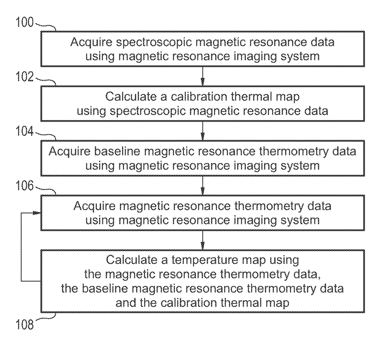
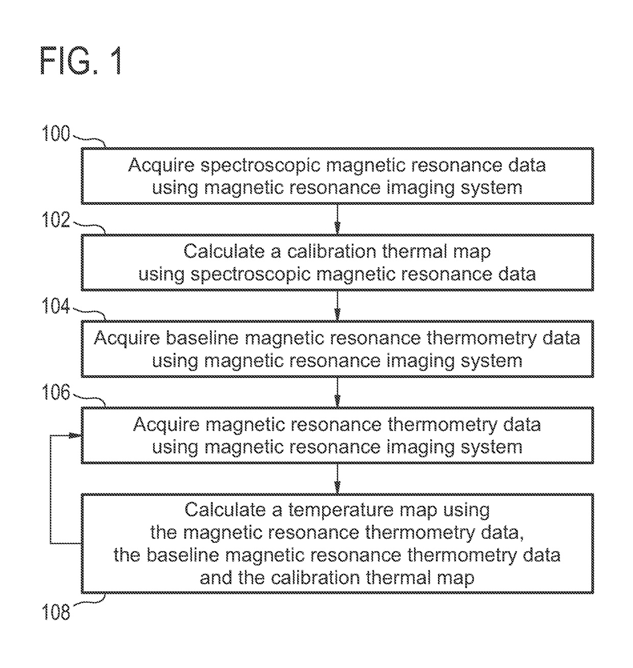
[0073]FIG. 1 shows a flow diagram which illustrates a method according to an embodiment of the invention. First in step 100 spectroscopic magnetic resonance data is acquired using a magnetic resonance imaging system. In step 102 a calibration thermal map is calculated using the spectroscopic magnetic resonance data. In step 104 baseline magnetic resonance thermometry data is acquired using the magnetic resonance imaging system. The baseline magnetic resonance thermometry data may be used to create a baseline set of measurements for comparison to the calibration thermal map. Next in step 106 magnetic resonance thermometry data is acquired using the magnetic resonance imaging system. In step 108 a temperature map is calculated using the magnetic resonance...


PUM


Abstract

A medical apparatus (300, 400, 500, 600) comprising a magnetic resonance imaging system (302). The medical apparatus further comprises a memory (332) storing machine readable instructions (352, 354, 356, 358, 470, 472, 474) for execution by a processor (326). Execution of the instructions causes the processor to acquire (100, 202) spectroscopic magnetic resonance data (334). Execution of the instructions further cause the processor to calculate (102, 204) a calibration thermal map (336) using the spectroscopic magnetic resonance data. Execution of the instructions further causes the processor to acquire (104, 206) baseline magnetic resonance thermometry data (338). Execution of the instructions further causes the processor to repeatedly acquire (106, 212) magnetic resonance thermometry data (340). Execution of the instructions further cause the processor to calculate (108, 214) a temperature map (351) using the magnetic resonance thermometry data, the calibration thermal map, and the baseline magnetic resonance thermometry data.

Description

CROSS-REFERENCE TO PRIOR APPLICATIONS[0001]This application is the U.S. National Phase application under 35 U.S.C. § 371 of International Application No. PCT / IB2012 / 050986, filed on Mar. 2, 2012, which claims the benefit of European Patent Application No. 11158606.1, filed on Mar. 17, 2011. These applications are hereby incorporated by reference herein.TECHNICAL FIELD[0002]The invention relates to magnetic resonance imaging, in particular the measurement of temperatures using magnetic resonance imaging.BACKGROUND OF THE INVENTION[0003]In recent years magnetic resonance thermometry has been coupled with various means of heating or cooling tissue for therapy. Measuring the effect of the tissue heating or cooling allows the guiding of the therapy and also the ability to assess the effect of a therapeutic treatment on a subject.[0004]In high-intensity focused ultrasound (HIFU) therapy, reliable real-time temperature monitoring using e.g. Magnetic Resonance Imaging (MRI) is necessary to ...

Claims


Application Information

Patent Timeline
Patent Type & Authority Patents(United States)
IPC IPC(8): A61B5/00A61B5/055G01R33/48A61B17/00A61B18/00A61N7/02A61N7/00A61B18/20A61B90/00A61B18/12A61B18/02A61B18/18
CPCG01R33/4804A61B5/055A61B18/02A61B18/12A61B18/1815A61B18/20G01R33/4814A61B2018/00791A61B2090/374A61N7/02A61N2007/0065G01R33/4808A61B2017/00084
Inventor KOHLER, MAX OSKARVAHALA, ERKKI TAPANINURMILAUKAS, KIRSI ILONA
Owner KONINKLIJKE PHILIPS ELECTRONICS NV
Why Eureka
Try Eureka