
Method for embedding a biological sample in a transparent matrix for analysis using single plane illumination microscopy

Active Publication Date: 2017-08-15
CARL ZEISS MICROSCOPY GMBH
View PDF2 Cites 2 Cited by
  • Summary
  • Abstract
  • Description
  • Claims
  • Application Information

AI Technical Summary

Benefits of technology

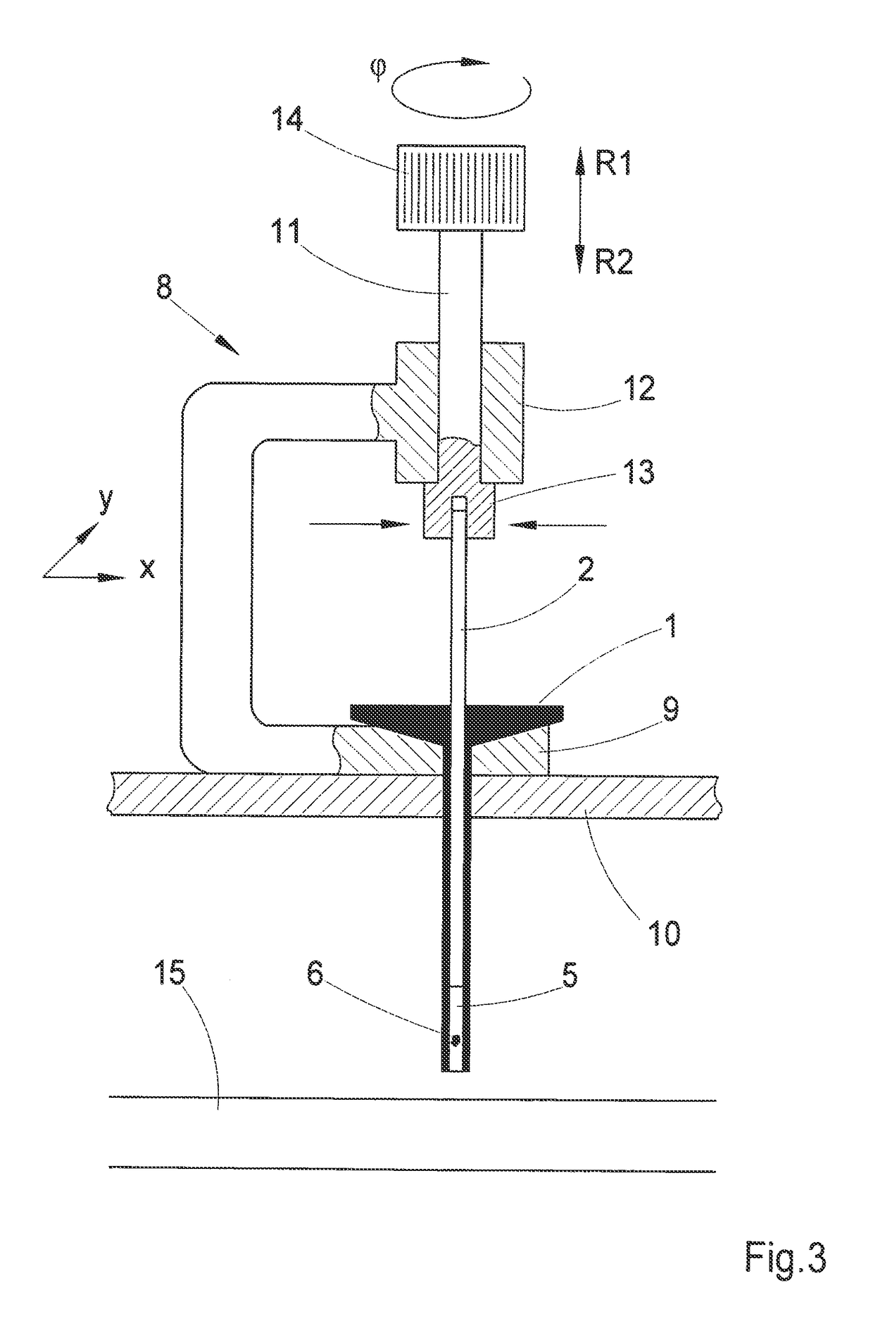
The present invention relates to a device called a capillary that can be used for removing liquid samples from a reservoir and delivering them to a larger container. The capillary has a small inner diameter and a piston that can be moved to create a suction effect to remove a portion of the liquid medium from the reservoir and push the liquid with the sample out of the capillary. The device can be designed to handle multiple capillaries simultaneously, allowing for efficient examination of samples. It is important that the piston comes into direct contact with the medium, without an air cushion, to facilitate the removal of viscous media such as gels and to prevent unwanted substances from entering via the air cushion. The capillary can be disconnected from the manipulating unit to immerse it in the medium and remove a portion of the medium using the piston. The technical effects of this invention include efficient and reliable removal of liquid samples for analysis and the ability to handle multiple samples simultaneously.

Problems solved by technology

At present, the positioning and alignment of the sample to be examined is carried out manually using commercially available equipment and is therefore extremely time-consuming and is accordingly only suitable for laboratory analyses of individual samples.

Method used


Image

  • Method for embedding a biological sample in a transparent matrix for analysis using single plane illumination microscopy
  • Method for embedding a biological sample in a transparent matrix for analysis using single plane illumination microscopy
  • Method for embedding a biological sample in a transparent matrix for analysis using single plane illumination microscopy

Examples


Embodiment Construction

[0068]It is to be understood that the figures and descriptions of the present invention have been simplified to illustrate elements that are relevant for a clear understanding of the present invention, while eliminating, for purposes of clarity, many other elements which are conventional in this art. Those of ordinary skill in the art will recognize that other elements are desirable for implementing the present invention. However, because such elements are well known in the art, and because they do not facilitate a better understanding of the present invention, a discussion of such elements is not provided herein.

[0069]The present invention will now be described in detail on the basis of exemplary embodiments.

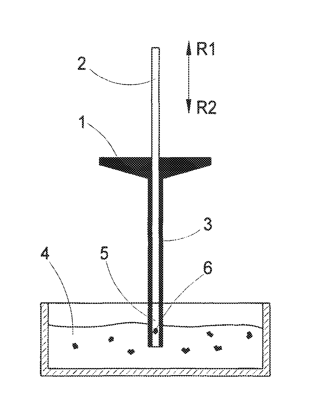
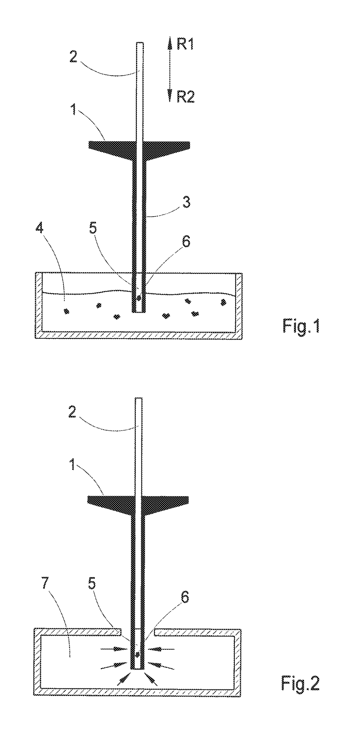
[0070]FIG. 1 shows a capillary 1 with a suction and delivery piston 2 which is guided in the interior so as to be displaceable in directions R1 and R2. Combinations of capillaries 1 and suction and delivery pistons 2 of this kind are known, per se, as pipettes and are used for ...


PUM


Abstract

The invention is directed to method for positioning and aligning a preferably biological sample in the detection area of the objective of a microscope arrangement. According to the invention, the method mentioned above has the following method steps: a sample is introduced into a transparent medium, preferably agarose gel, which is initially liquid; the medium is changed from the liquid state to the solid state, wherein the sample is fixated within the medium, but the transparency of the medium is retained; the solidified medium is positioned in the microscope arrangement in such a way that the sample enclosed therein is situated in the detection area of the objective. Further, a device is proposed for positioning and aligning a preferably biological sample in the detection area of the objective of a microscope arrangement.

Description

[0001]The present application is a continuation of U.S. patent application Ser. No. 12 / 997,113 filed on Dec. 9, 2010, which claims priority from PCT Patent Application No. PCT / EP2009 / 004081 filed on Jun. 6, 2009, which claims priority from German Patent Application No. DE 10 2008 027 784.3 filed on Jun. 11, 2008, the disclosures of which are incorporated herein by reference in their entirety.BACKGROUND OF THE INVENTION[0002]1. Field of the Invention[0003]The invention is directed to a method and a device for positioning and aligning a preferably biological sample in the detection area of the objective in a microscope arrangement.[0004]The method according to the invention can be applied particularly in connection with single plane illumination microscopy (SPIM). This microscopy method is a special method of widefield microscopy by which image data for a three-dimensional image of the sample are obtained on the basis of optical sections through different planes of the sample.[0005]2....

Claims


Application Information

Patent Timeline
Patent Type & Authority Patents(United States)
IPC IPC(8): G01N1/28G02B21/32G02B21/34
CPCG01N1/28G02B21/32G02B21/34
Inventor HELLMICH, WIBKERADT, BENNOLIPPERT, HELMUTSELCHOW, OLAFWOLF, UWEHAESE, JUERGEN
Owner CARL ZEISS MICROSCOPY GMBH
Learn More