
Cross sectional depth composition generation utilizing scanning electron microscopy

a scanning electron microscope and composition technology, applied in the direction of material analysis using wave/particle radiation, material analysis by measuring secondary emission, instruments, etc., can solve the problems of too small deconstructibility or too fragile cross-sections

Inactive Publication Date: 2017-07-11
IBM CORP
View PDF10 Cites 4 Cited by
  • Summary
  • Abstract
  • Description
  • Claims
  • Application Information

AI Technical Summary

Problems solved by technology

In many instances, the specimens to be cross-sectioned are too fragile or too small to deconstruct or cross-section, or the specimens need to be preserved for other analyses where non-destructive testing is needed.

Method used


Image

  • Cross sectional depth composition generation utilizing scanning electron microscopy
  • Cross sectional depth composition generation utilizing scanning electron microscopy
  • Cross sectional depth composition generation utilizing scanning electron microscopy

Examples


Embodiment Construction

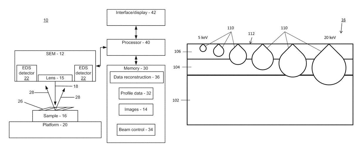
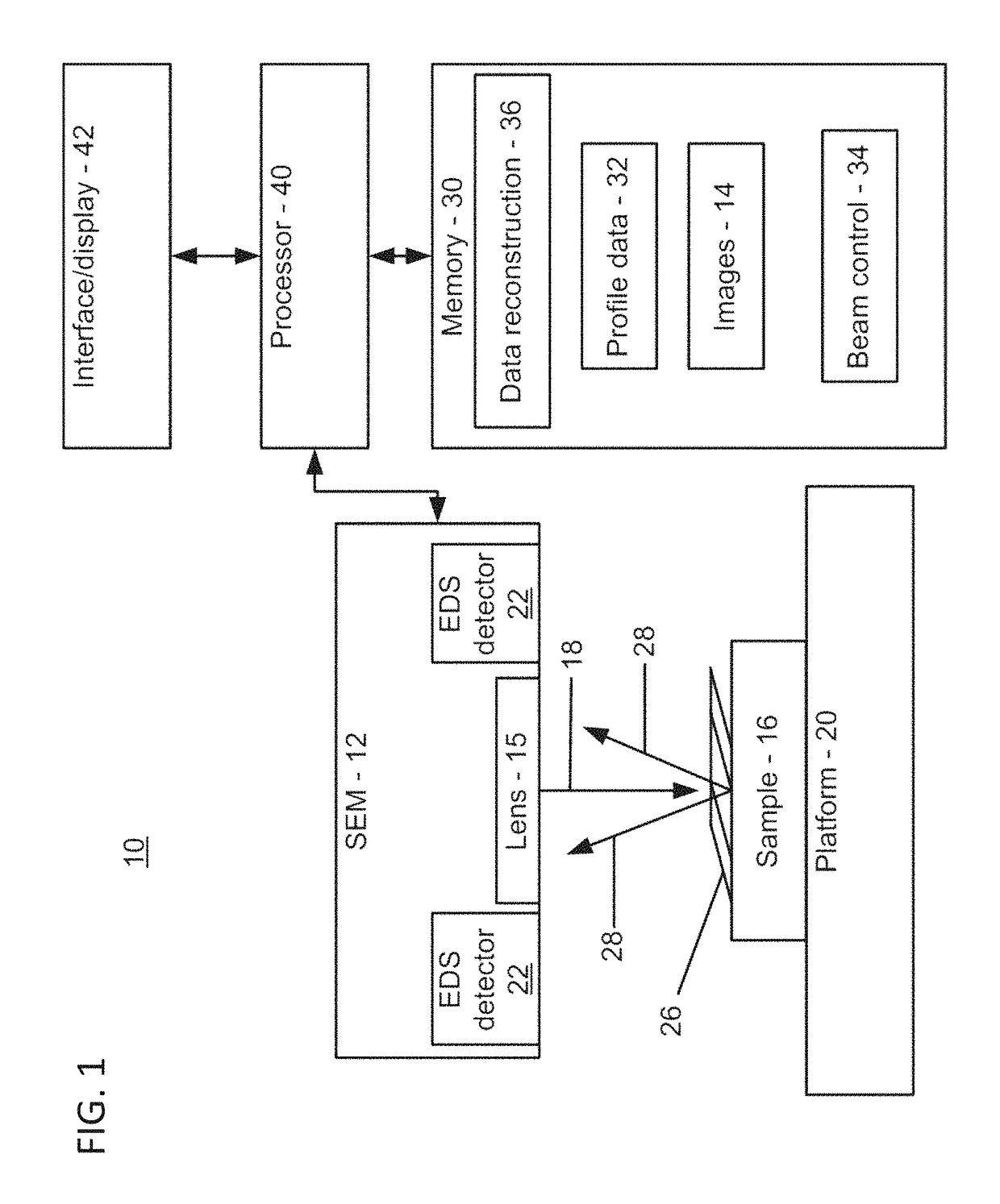
[0017]In accordance with the present principles, a scanning electron microscope (SEM) is employed to analyze materials to generate a cross-sectional depth composition into a surface of a sample. This is especially useful for small samples that are extremely difficult to manually cross-section and for specimens where non-destructive testing is needed. In one embodiment, a SEM creates a cross-sectional view into the sample by varying a beam voltage of the SEM. A cross-sectional depth composition of a surface is determined by performing a plurality of point scans using a SEM at various beam energy levels and then comparing electron dispersive X-ray spectroscopy (EDS) results at those various energy levels to create a depth composition.

[0018]In one illustrative method, beam penetration (which has a tear drop shape) is measured at various beam energy levels. As the beam energy is progressively increased or decreased, the composition that is measured by EDS detectors changes. These change...


PUM

PropertyMeasurementUnit
energyaaaaaaaaaa
energy levelaaaaaaaaaa

Abstract

A method for generating cross-sectional profiles using a scanning electron microscope (SEM) includes scanning a sample with an electron beam to gather an energy-dispersive X-ray spectroscopy (EDS) spectrum for an energy level to determine element composition across an area of interest. A mesh is generated to locate positions where a depth profile will be taken. EDS spectra are gathered for energy levels at mesh locations. A number of layers of the sample are determined by distinguishing differences in chemical composition between depths as beam energies are stepped through. A depth profile is generated for the area of interest by compiling the number of layers and the element composition across the mesh.

Description

BACKGROUND[0001]Technical Field[0002]The present invention relates to characterizing layers of an object, and more particularly to systems and methods for generating cross-sectional composition and structural information using a scanning electron microscope (SEM).[0003]Description of the Related Art[0004]A scanning electron microscope (SEM) produces images of a sample or specimen by scanning with a focused beam of electrons. The electrons interact with atoms in the sample and produce signals that include information about a sample's surface topography and composition. An electron beam is scanned, and its position is combined with a detected signal to produce an image. SEM can achieve resolutions of better than 1 nanometer.[0005]The electron beam may have an energy ranging from 0.2 keV to 40 keV and is focused by one or more condenser lenses. The beam scans in a raster pattern over an area of a sample. SEM can provide several items of data at each pixel. One example includes energy-d...

Claims


Application Information

Patent Timeline
Patent Type & Authority Patents(United States)
IPC IPC(8): G01N23/225G01B15/00G01B15/04
CPCG01N23/2252G01B15/00G01B15/04G01N2223/079G01N2223/418G01B15/02G01N23/22H01J2237/221H01J2237/24578H01J2237/24585H01J2237/248H01J2237/2807G01N2223/304G01N2223/633H01J37/222H01J37/28H01J2237/2815
Inventor CAMPBELL, ERIC J.CZAPLEWSKI, SARAH K.
Owner IBM CORP
Learn More