High surface area substrate for cell culture
a cell culture and substrate technology, applied in the field of cell culture microcarriers, can solve the problems affecting the ability and nature of the binding of certain cells to the microcarrier, and achieve the effects of not prone to delamination, easy to tune, and limit the risk of pathogen contamination
- Summary
- Abstract
- Description
- Claims
- Application Information
AI Technical Summary
Benefits of technology
Problems solved by technology
Method used
Image
Examples
example 1
Vitronectin (VN) Conjugation to Polystyrene Beads
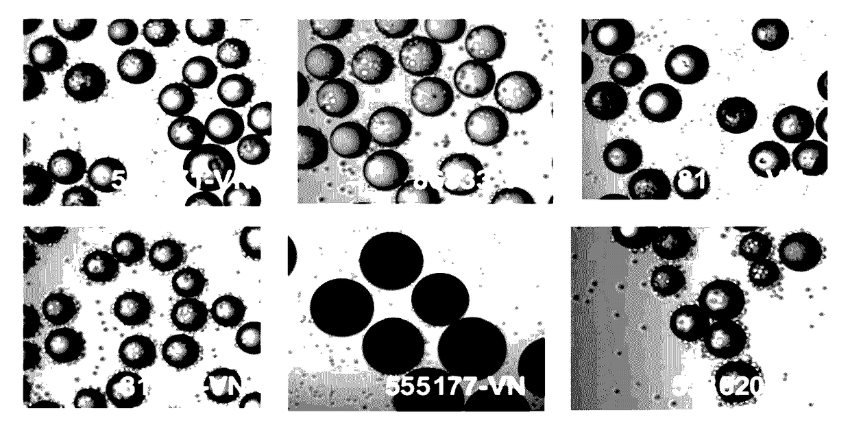
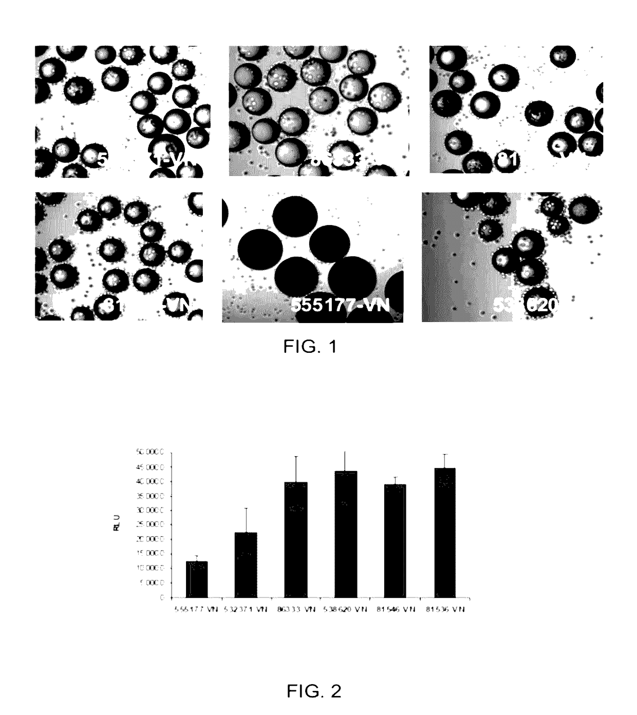
[0073]100 mg of the COOH functionalized polystyrene microspheres (see Table 1 below for linkers used) were transferred to a 2 mL centrifuge tube, 0.4 g of EDC and 0.1 g of NHS was dissolved in 20 mL of DMF to prepare a stock solution. An aliquot of the solution was added and then allowed to mix on an orbital shaker for 60 min. The solution was aspirated, rinsed once with DMF, aspirated and then an aliquot of the following sequence peptide solution [Ac-Lys-Gly-Gly-Pro-Gln-Val-Thr-Arg-Gly-Asp-Val-Phe-Thr-Met-Pro-NH2] (10 mM in borate buffer, pH 9.2, 0.25% Rhodamine peptide spiked) was added and allowed mix for 60 mM. The peptide solution was removed by aspiration and the spheres were treated with 1.5 mL of 1M ethanolamine pH 8 for 10 min followed by washing with PBS (1.5 mL×5), 1% SDS (1×1.5 mL×1.5 min), DI Water (1.5 mL×5), ethanol (1.5 mL×5) and dried under a gentle stream of nitrogen. Vitronectin conjugated to beads, identified in Ta...
example 2
Cell Culture with HT1080 Cells
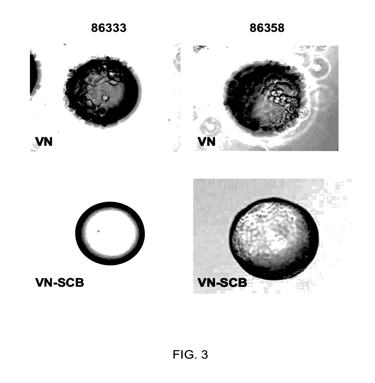
[0075]HT1080 cells (ATCC) were routinely expanded on TCT flasks (Corning, N.Y.) in Iscove's Modified Dulbecco's Medium (IMDM) with 10% Fetal Bovine Serum (FBS). For cell adhesion assay, cells were trypsinized and allowed to recover in Iscove's Modified Dulbecco's Medium (IMDM) with 10% Fetal Bovine Serum (FBS) for 30 minutes at 37° C., 5% CO2. After recovery, the cells were washed and resuspended in 0.1% Bovine Serum Albumin (BSA) in IMDM. Approximately 10 mg of peptide derivatized microcarriers were transferred to a 2 mL centrifuge tube and blocked with 2 mL of 1% BSA in D-PBS for 1 hr at room temperature. The microcarriers were then aspiration washed with 2 mL of D-PBS and incubated with 2 mL of 0.1% BSA in IMDM prior to cell seeding. 2 mL of re-suspended cells (200 000 cells / well) were placed in several wells of a 24 well Corning Ultra low attachment microplate. To each cell seeded well was added approximately 10 mg of peptide coated microcarriers an...
example 3
Cell Culture with ReNcell VM Cells
[0077]ReNcell VM cells, human neural progenitor cells, from Millipore (Temecula, Calif.) were routinely expanded on laminin coated T75 cm2 tissue culture flasks (Corning, N.Y.) in ReNcell NSC Maintenance Medium (Millipore, Temecula, Calif.) containing 20 ng / mL FGF-2 and 20 ng / mL EGF (Millipore, Temecula, Calif.). For maintenance and growth of undifferentiated cells, the medium was changed every day. All cells in culture were maintained at 37° C. in a humidified atmosphere of 95% air / 5% CO2. Cells were passaged once a week using Accutase™ (Millipore, Temecula, Calif.). For cell adhesion assay, undifferentiated cells were detached by treatment with accutase and allowed to recover in ReNcell NSC Maintenance Medium for 30 minutes at 37° C., 5% CO2. After recovery, the cells were washed and resuspended in 0.1% Bovine Serum Albumin (BSA) in ReNcell NSC Maintenance Medium. Approximately 10 mg of peptide derivatized microcarriers were transferred to a 2 mL ...
PUM
| Property | Measurement | Unit |
|---|---|---|
| density | aaaaa | aaaaa |
| pH | aaaaa | aaaaa |
| pH | aaaaa | aaaaa |
Abstract
Description
Claims
Application Information
 - R&D
- Intellectual Property
- Life Sciences
- Materials
- Tech Scout
- Unparalleled Data Quality
- Higher Quality Content
- 60% Fewer Hallucinations
Browse by: Latest US Patents, China's latest patents, Technical Efficacy Thesaurus, Application Domain, Technology Topic, Popular Technical Reports.
© 2025 PatSnap. All rights reserved.Legal|Privacy policy|Modern Slavery Act Transparency Statement|Sitemap|About US| Contact US: help@patsnap.com


