Eureka AIR delivers breakthrough ideas for toughest innovation challenges, trusted by R&D personnel around the world.

High Surface Area Substrate for Cell Culture

a cell culture and substrate technology, applied in the field of cell culture microcarriers, can solve the problems affecting the ability and nature of the binding of certain cells to the microcarrier, and achieve the effects of not prone to delamination, easy to tune, and limit the risk of pathogen contamination

Active Publication Date: 2012-03-08
CORNING INC
View PDF3 Cites 8 Cited by
  • Summary
  • Abstract
  • Description
  • Claims
  • Application Information

AI Technical Summary

Benefits of technology

[0007]Among other things, the present disclosure describes synthetic, chemically-defined microcarriers that may provide robust and selective binding of anchorage-dependent cells. The microcarriers may be formed from conjugation of a polypeptide to a carboxylic acid group attached to the surface of a polystyrene microcarrier base. The polypeptide may contain an RGD amino acid sequence for mediating bio-specific cell adhesion. As described herein, such microcarriers allow for selective adhesion of cells via the polypeptide with little or no non-selective binding. Accordingly, the microcarriers described herein may be used to culture anchorage dependent cells in a controlled manner. As also described herein, such microcarriers allow the specific attachment and cell growth of pluripotent stem cells, including human embryonic stem cells (hESC).
[0008]In various embodiments, after grafting the polypeptides to carboxylic acid groups attached to the microcarrier base, the remaining non-peptide conjugated carboxylic acid groups, or a portion thereof, are blocked via derivation with a low molecular weight amine monamine, such as ethanolamine. Such blocking converts highly hydrophilic carboxylic acid groups to less hydrophilic amide group, which may affect the ability and nature of the binding of certain cells to the microcarrier. Accordingly, the hydrophilic characteristics of the microcarrier can be readily tuned, as desired, to impact cell behavior, due to the percentage of COOH groups blocked and the nature of the monoamine used for blocking.
[0009]One or more embodiments of the microcarriers or methods described herein provide one or more advantages over prior microcarriers, methods for preparing microcarriers, or methods for culturing cells with microcarriers. For example, the microcarriers described herein are devoid of animal derived materials, which limits the risk of pathogen contamination. This is particularly relevant when the cultured cells are dedicated to cell-therapies. The microcarriers may be monolithic and not coated, and thus are not prone to delamination. Due to low levels of non-specific binding to the microcarrier base, bioselective cell attachment through receptor binding to polypeptides grafted to the microcarrier can occur. Such highly bioselective attachment may be beneficial for culturing primary cells, hepatocytes, and other cells, such as stem cells including embryonic stem cells. In addition to ready tuning of the hydrophilicity of the surface of the microcarrier, the polypeptide density can be tuned by derivation of the carboxylic acid groups of the microcarrier based. These and other advantages will be readily understood from the following detailed descriptions when read in conjunction with the accompanying drawings.

Problems solved by technology

Such blocking converts highly hydrophilic carboxylic acid groups to less hydrophilic amide group, which may affect the ability and nature of the binding of certain cells to the microcarrier.

Method used


Image

  • High Surface Area Substrate for Cell Culture
  • High Surface Area Substrate for Cell Culture
  • High Surface Area Substrate for Cell Culture

Examples


example 1

Vitronectin (VN) Conjugation to Polystyrene Beads

[0073]100 mg of the COOH functionalized polystyrene microspheres (see Table 1 below for linkers used) were transferred to a 2 mL centrifuge tube, 0.4 g of EDC and 0.1 g of NHS was issolved in 20 mL of DMF to prepare a stock solution. An aliquot of the solution was added and then allowed to mix on an orbital shaker for 60 min. The solution was aspirated, rinsed once with DMF, aspirated and then an aliquot of the following sequence peptide solution [Ac-Lys-Gly-Gly-Pro-Gln-Val-Thr-Arg-Gly-Asp-Val-Phe-Thr-Met-Pro-NH2] (10 mM in borate buffer, pH 9.2, 0.25% Rhodamine peptide spiked) was added and allowed mix for 60 mM. The peptide solution was removed by aspiration and the spheres were treated with 1.5 mL of 1 M ethanolamine pH 8 for 10 min followed by washing with PBS (1.5 mL×5), 1% SDS (1×1.5 mL×1.5 min), DI Water (1.5 mL×5), ethanol (1.5 mL×5) and dried under a gentle stream of nitrogen. Vitronectin conjugated to beads, identified in Ta...

example 2

Cell Culture with HT1080 Cells

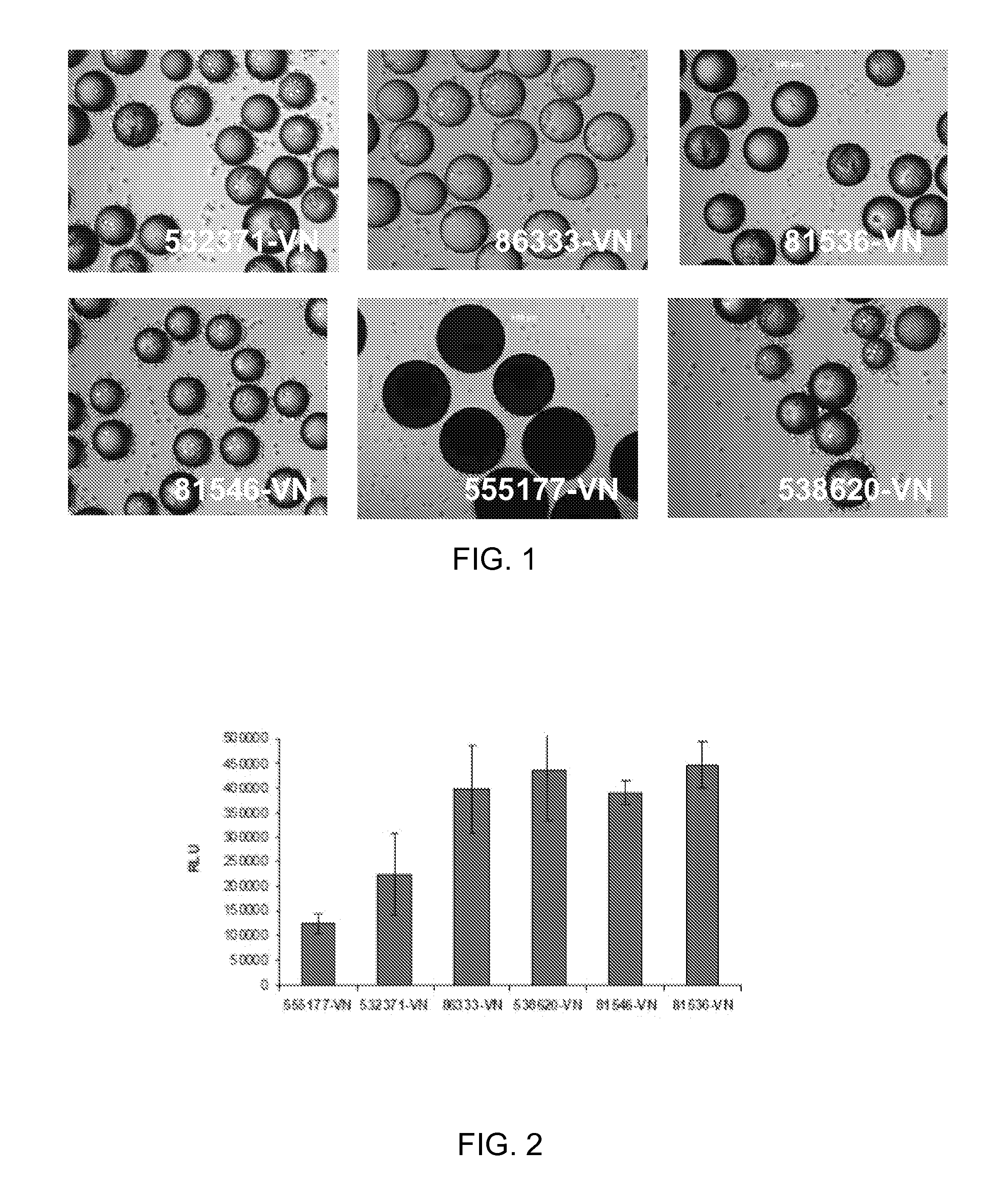
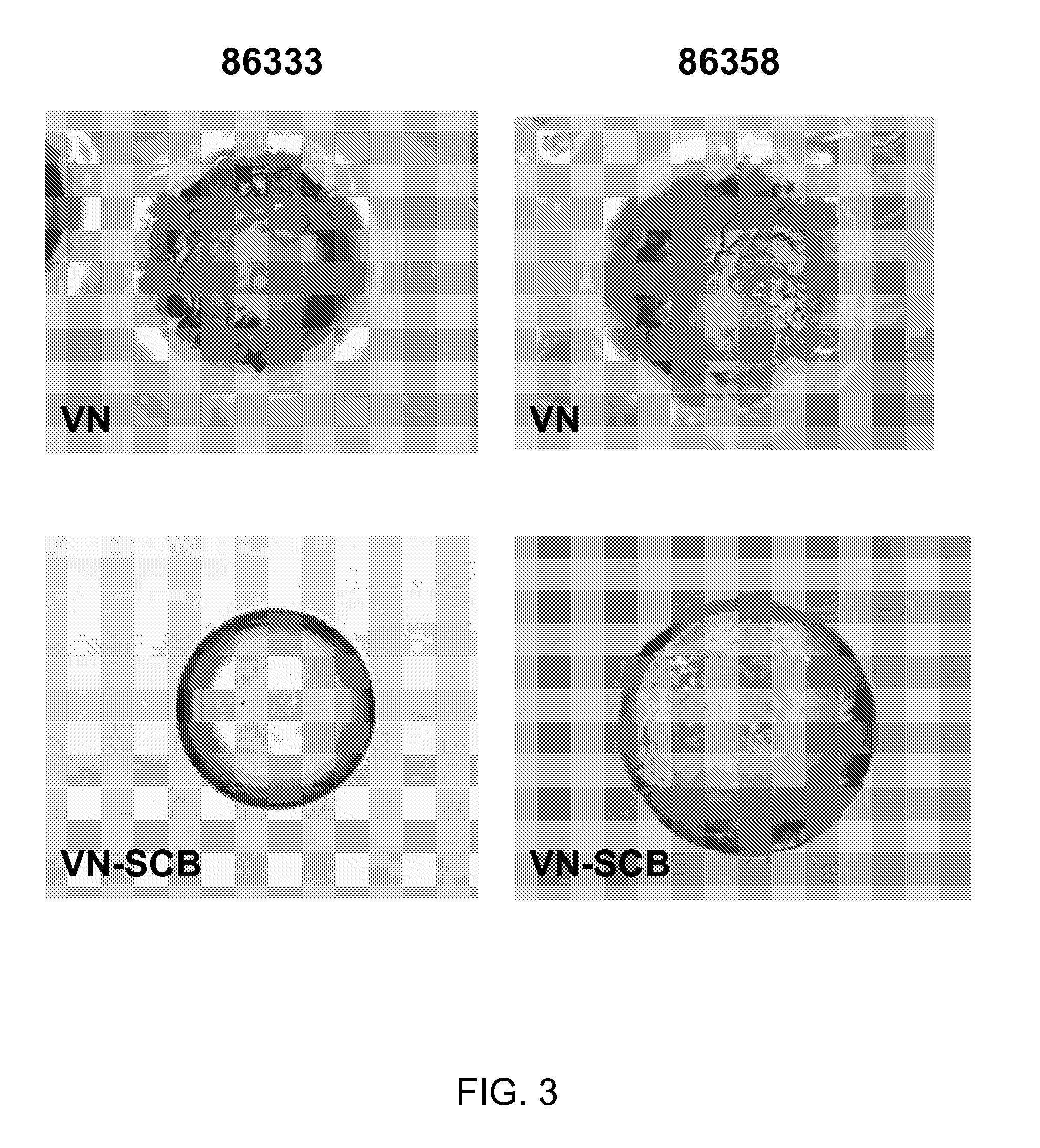
[0074]HT1080 cells (ATCC) were routinely expanded on TCT flasks (Corning, N.Y.) in Iscove's Modified Dulbecco's Medium (IMDM) with 10% Fetal Bovine Serum (FBS). For cell adhesion assay, cells were trypsinized and allowed to recover in Iscove's Modified Dulbecco's Medium (IMDM) with 10% Fetal Bovine Serum (FBS) for 30 minutes at 37° C., 5% CO2. After recovery, the cells were washed and resuspended in 0.1% Bovine Serum Albumin (BSA) in IMDM. Approximately 10 mg of peptide derivatized microcarriers were transferred to a 2 mL centrifuge tube and blocked with 2 mL of 1% BSA in D-PBS for 1 hr at room temperature. The microcarriers were then aspiration washed with 2 mL of D-PBS and incubated with 2 mL of 0.1% BSA in IMDM prior to cell seeding. 2 mL of re-suspended cells (200 000 cells / well) were placed in several wells of a 24 well Corning Ultra low attachment microplate. To each cell seeded well was added approximately 10 mg of peptide coated microcarriers an...

example 3

Cell Culture with ReNcell VM Cells

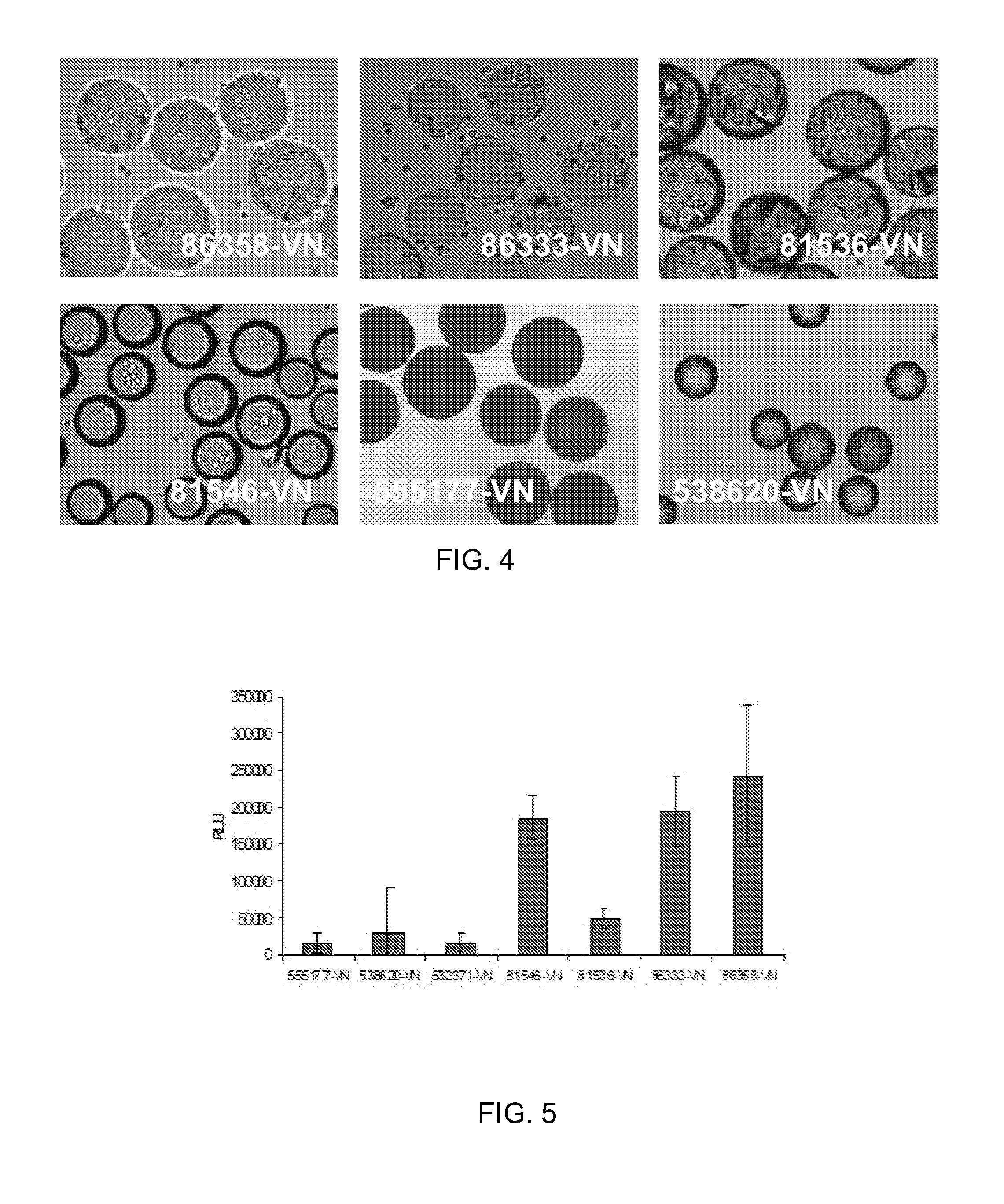
[0076]ReNcell VM cells, human neural progenitor cells, from Millipore (Temecula, Calif.) were routinely expanded on laminin coated T75 cm2 tissue culture flasks (Corning, N.Y.) in ReNcell NSC Maintenance Medium (Millipore, Temecula, Calif.) containing 20 ng / mL FGF-2 and 20 ng / mL EGF (Millipore, Temecula, Calif.). For maintenance and growth of undifferentiated cells, the medium was changed every day. All cells in culture were maintained at 37° C. in a humidified atmosphere of 95% air / 5% CO2. Cells were passaged once a week using Accutase™ (Millipore, Temecula, Calif.). For cell adhesion assay, undifferentiated cells were detached by treatment with accutase and allowed to recover in ReNcell NSC Maintenance Medium for 30 minutes at 37° C., 5% CO2. After recovery, the cells were washed and resuspended in 0.1% Bovine Serum Albumin (BSA) in ReNcell NSC Maintenance Medium. Approximately 10 mg of peptide derivatized microcarriers were transferred to a 2 mL ...


PUM

PropertyMeasurementUnit
Molalityaaaaaaaaaa
Molalityaaaaaaaaaa
Sizeaaaaaaaaaa

Abstract

A cell culture microcarrier includes (1) a polystyrene microcarrier base having a remnant of a carboxylic acid group, and (ii) a polypeptide conjugated to the base via the remnant of the carboxylic acid group. The polypeptide may contain a cell adhesive sequence, such as RGD. Cells cultured with such microcarriers exhibit peptide-specific binding to the microcarriers.

Description

CROSS-REFERENCE TO RELATED APPLICATIONS[0001]This application claims the benefit of priority under 35 U.S.C. §119(e) of U.S. Provisional Application Ser. No. 61 / 308,101 filed on Feb. 25, 2010 which claims the benefit of priority under 35 U.S.C. §119(e) of U.S. Provisional Application Ser. No. 61 / 229,114 filed on Jul. 28, 2009.FIELD[0002]The present disclosure relates to cell culture microcarriers, and more particularly to synthetic, chemically-defined microcarriers.SEQUENCE LISTING[0003]This application contains a Sequence Listing electronically submitted via EFS-Web to the United States Patent and Trademark Office as text filed named “SP10-044_SEQ_LIST_ST25.txt” having a size of 1 kb and created on Jul. 28, 2010. Due to the electronic filing of the Sequence Listing, the electronically submitted Sequence Listing serves as both the paper copy required by 37 CFR §1.821(c) and the CRF required by §1.821(e). The information contained in the Sequence Listing is hereby incorporated herein...

Claims


Application Information

Patent Timeline
IPC IPC(8): C12N5/071C08F12/08C12N1/20C12N5/0735C12N5/04C12N5/078C12N5/09C12N5/0797C08F8/30C12N1/16
CPCC12N5/0075C12N2533/50C12N2533/30C12N5/0606
Inventor FABIAN, MICHELLE DAWNMYERS, TIMOTHY EDWARDSNYDER, KYLE PATRICKVERRIER, FLORENCE
Owner CORNING INC
Why Eureka
Eureka Blog
Learn More