Surface electrode design that can be left in place during MR imaging

a surface electrode and imaging technology, applied in the direction of reradiation, magnetic variable regulation, sensors, etc., can solve the problems of reducing image quality, induced current can be a burn risk for patients, and the surface electrode is not designed for use in an mr, so as to reduce the risk of heating, reduce the effect of eddy current and their impact on image quality, and reduce the surface area

Active Publication Date: 2016-08-16
DEERFIELD IMAGING INC
View PDF26 Cites 40 Cited by
  • Summary
  • Abstract
  • Description
  • Claims
  • Application Information

AI Technical Summary

Benefits of technology

[0019]and reducing artifacts in the generated image by dividing the layer into separate side by side sections where the separation into the side by side sections is arranged to reduce eddy currents which are induced in the surface electrodes when they are exposed to variations in the magnetic field and to RF pulses;
[0042]This invention relates to the system disclosed in PCT Published Application 2009 / 0306494 of the present applicant published Dec. 10 2009, the disclosure of which is incorporated herein by reference. The arrangement disclosed herein enables the above system by improving the workflow between X-ray fluoro-guided catheter interventions and post-intervention MR imaging. The novelty exists in a cut pattern that reduces the eddy current's effect on image quality as well as the type of connector and cable such that the connection / disconnection can be made quickly; that the length of cable, if any, remaining attached to the electrodes does not pose a burn risk to the patient, and integrating these elements (connector / cuts) without degrading the information that the electrode is originally intended to provide.

Problems solved by technology

However, surface electrodes are not designed for use in an MR.
The patch component is a conductive surface and therefore susceptible to eddy currents from the scanner magnetic gradients and RF pulses.
These currents degrade image quality.
The wire acts as an antenna with respect to the RF pulses and the induced current can be a burn risk for the patient.
These issues, MR image degradation and safety risk, generally require that the surface electrodes must be removed from the patient before MR imaging occurs.
However, this is not the optimal workflow as the MR post-interventional imaging may reveal a need for further immediate treatment.
This extra effort also occurs for the above system but may require that the impedance map of the heart be re-acquired.

Method used


Image

  • Surface electrode design that can be left in place during MR imaging
  • Surface electrode design that can be left in place during MR imaging
  • Surface electrode design that can be left in place during MR imaging

Examples


Embodiment Construction

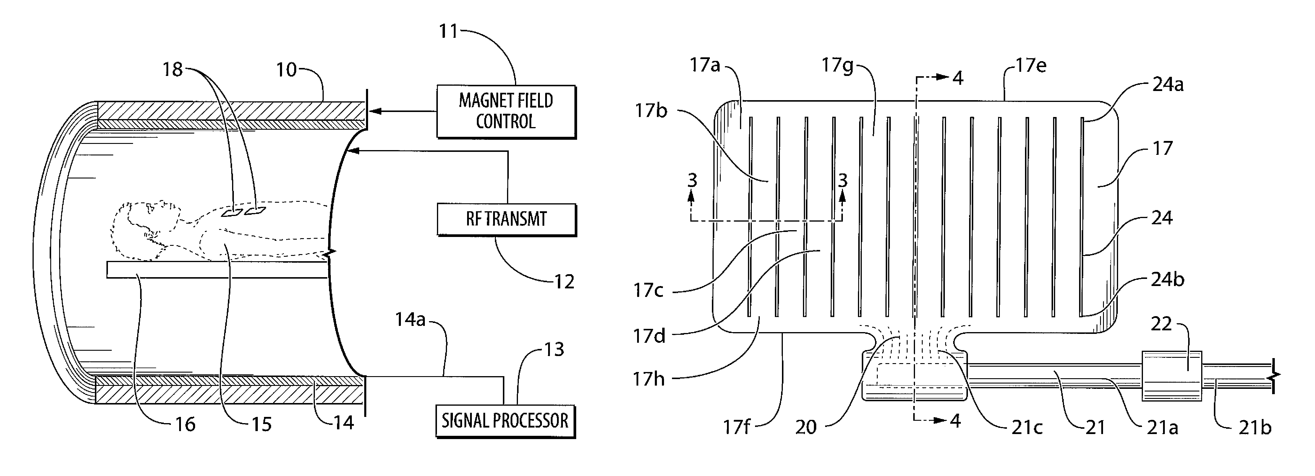
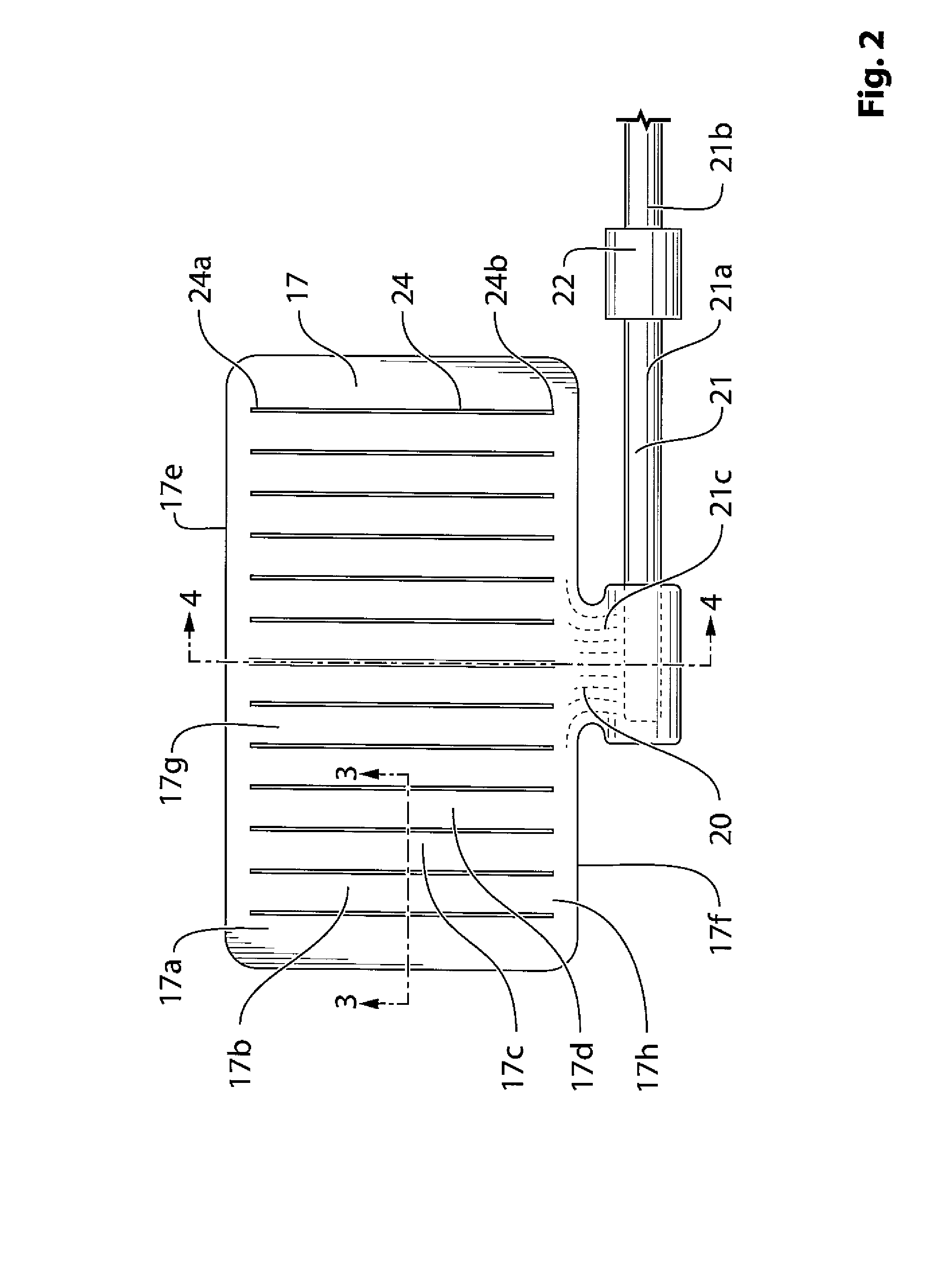
[0052]The apparatus for MR imaging of a subject includes a conventional cylindrical MR magnet 10 operable by a field control system to generate a variable magnetic field to be applied to the subject.

[0053]The MR system includes an RF transmit arrangement 12 for generating RF pulses in a transmit stage to be applied to the subject to be imaged and a receive arrangement for acquiring the MR signal in a receive stage with a signal processing system 13 for receiving the MR signal for carrying out signal processing by which an image is generated. A subject 15 is carried on a table 16 located within the imaging space of the magnet 10. As is well known, the subject generates an MR signal in response to the magnetic field and the RF signal applied which is detected and processed to generate an image. The arrangement is well known and a suitable system is available from Siemens.

[0054]Typically the magnet 10 carries an RF coil known as a body coil 14 which is mounted on the cylindrical magnet...


PUM


Abstract

For use in MR imaging of a patient a plurality of surface electrodes such as ECG or defibrillator are provided for obtaining electrical signals for determining electrical activity within the body of the patient and remain in place during the MR imaging. The surface electrodes include a quick disconnect wire for carrying the signals to a signal processing system to be removed during the MR imaging to prevent heating. Each electrode comprises a conductive layer divided by slits into separate side by side sections to reduce eddy currents which are induced in the surface electrodes when they are exposed to variations in the magnetic field. The sections all are connected through the layer to the conductive location to allow the signal therefrom to be connected to the communication conductor.

Description

[0001]This invention relates to a surface electrode design that can be left in place during MR imaging.BACKGROUND OF THE INVENTION[0002]Surface electrodes are wired electrical connections commonly used to measure biological signals. These electrodes support patient monitoring by measuring electrical signals related to brain function (EEG) and heart activity (ECG) etc. Typically, many electrodes are distributed on the surface of the head or body to collect, in aggregate, diagnostic information. For example, 12-lead ECG can identify characteristic information about an ST-segment elevated myocardial infarction, a common type of heart attack.[0003]Surface electrodes are also used to track invasive devices such as the St Jude Medical ENSITE NAVX (trademark) catheter tracking system. This device uses three pairs of surface electrodes positioned to be create roughly perpendicular electric fields through the body. A similar array of electrodes is used by Biosense Webster CARTO3 system. Thes...

Claims


Application Information

Patent Timeline
Patent Type & Authority Patents(United States)
IPC IPC(8): A61B5/0424A61B5/055G01R33/567G01R33/28A61B5/0408A61B5/276A61B5/296
CPCA61B5/04087G01R33/288G01R33/5673A61B5/0424A61B5/055A61B2562/0223A61B5/259A61B5/276
Inventor LANG, MICHAELPETROPOLOUS, LABROSALEXIUK, MARK
Owner DEERFIELD IMAGING INC
Why Eureka
Try Eureka