
Vascular wound closing apparatus and method

a wound and wound technology, applied in the field of wound closure apparatus and methods, can solve the problems of increasing hospital stays, affecting morbidity, poor wound closure, etc., and achieve the effect of reducing patient blood pressure and flow

Active Publication Date: 2016-02-16
ENSITEVASCULAR LLC
View PDF47 Cites 2 Cited by
  • Summary
  • Abstract
  • Description
  • Claims
  • Application Information

AI Technical Summary

Benefits of technology

The patent describes a device designed to reduce a patient's blood pressure and flow during a medical procedure. It achieves this by using a force-exerting assembly that applies a constant and symmetrically distributed force through a force-transmitting surface. This design helps to control the blood flow and can accommodate different patient needs, such as sheath sizes and blood chemistries. Overall, the device helps to improve the precision and safety of the medical procedure.

Problems solved by technology

Poorly executed wound closures may give rise to complications which are costly, increase hospital stays and affect morbidity.
For example, inadequate hemostasis can lead to significant blood loss, patient discomfort, vessel occlusion, thrombosis, formation of arteriovenous fistula, and pseudoaneurysm requiring surgical intervention and / or steps to avoid infections.
However, in actual practice, there are a number of significant problems.
For example, manual pressure that is too firm does not allow sufficient clotting factors to accumulate at the arteriotomy.
Even more important, the person exerting manual pressure can tire during the 30-minute holding time, or the fingers may move or may not be placed properly.
The person may also temporarily stop the application of pressure to examine the wound, causing a disruption of the maturing clot.
Finally, different body types present different manual pressure issues, e.g., if the panniculus intrudes on the person's left hand, pressure variations may be induced as the patient breathes and the panniculus moves.
These combined techniques have many of the same problems as straightforward manual pressure closures, and the external devices may be difficult to deploy on obese patients.
Thus, while manual procedures are of long standing, they are deficient in that they can be tiring, require careful training, and represent inefficient use of the time of valuable medical personnel.
Drawbacks of these procedures include a maximum French size of 8 Fr and the fact that the anchor and collagen plug must be left in the body for up to 90 days.
Suture-mediated intra-arterial anchor techniques have also been used, but these are deficient in that the sutures remain in the body until absorbed, and nonetheless require that the anchor and plug be left in the body for an extended period.
In response to these problems, various specialized vascular closure devices (VCDs) have been proposed, such as the device disclosed in U.S. Pat. No. 5,307,811 and commercialized under the designation “FemoStop.” While these and other VCDs have achieved widespread use, no prior VCD has fully solved the problems inherent in wound closures.
A further complication is that the heart is beating, making the pressure (sum of internal and external pressure) variable.
If there were no flow restriction, the arteriotomy would leak, resulting in a hematoma.

Method used


Image

  • Vascular wound closing apparatus and method
  • Vascular wound closing apparatus and method
  • Vascular wound closing apparatus and method

Examples


Embodiment Construction

The Preferred Wound Closure Apparatus of FIGS. 1-23

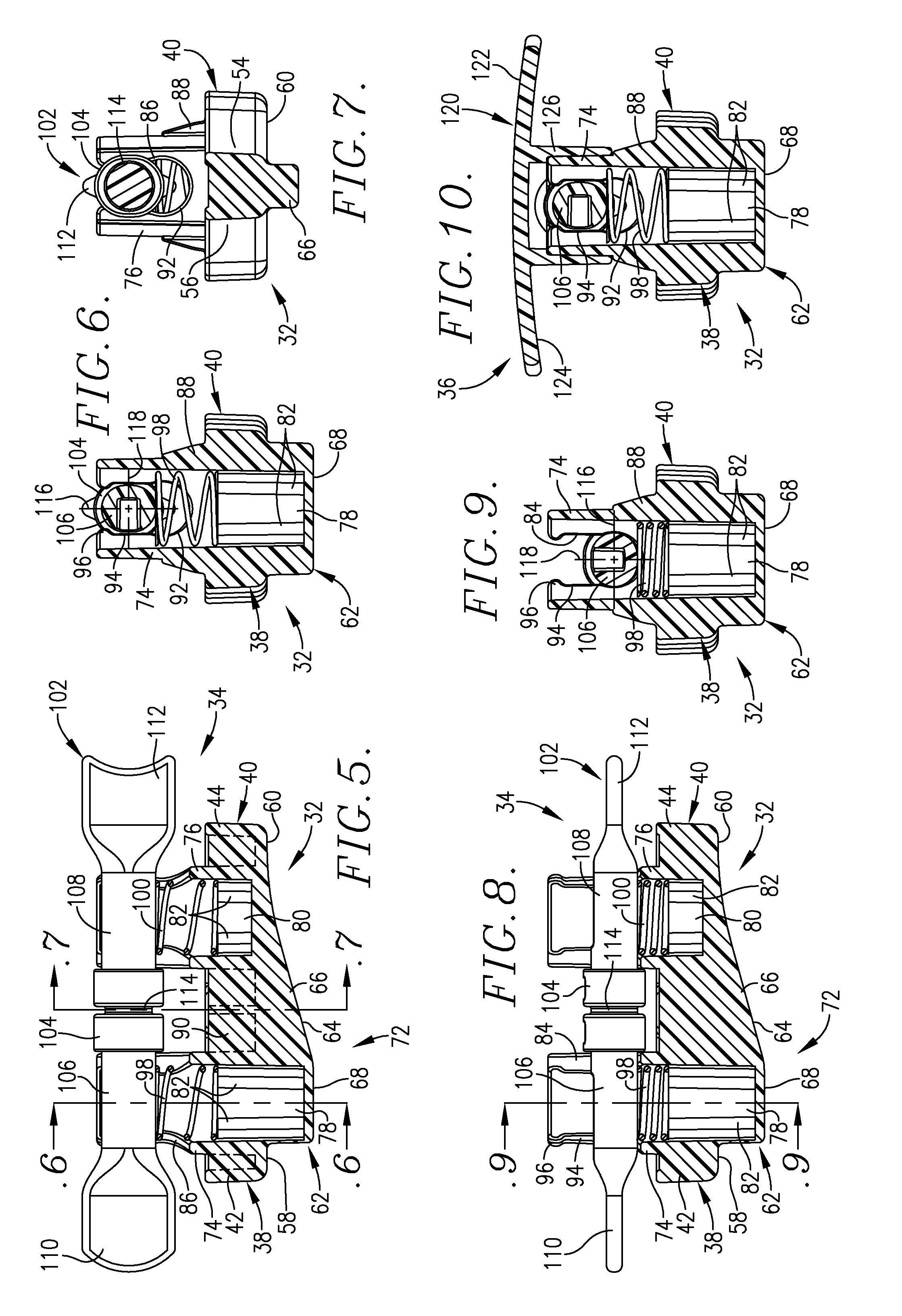
[0063]Turning now to the drawings, apparatus 30 operable to close a wound in a patient's tissue is illustrated in FIGS. 1-4. The apparatus 30 is particularly designed for closure of wounds attendant to an endovascular (i.e., arterial or venous) intervention involving, e.g., a femoral artery puncture where the wound presents an insertion site, an elongated, obliquely oriented tract extending into the patient's tissue and communicating with the insertion site and an arteriotomy. Broadly speaking, the apparatus 30 includes a force-transmitting body 32 having a force-exerting assembly 34 together with a removable cover or “hat”36.

[0064]As used herein, terms such as “upper” and “lower,”“top and “bottom,” and “downwardly” and “upwardly” and the like are used for convenience and because of the fact that the apparatus 30 is normally positioned in an upright orientation on a patient with the cover 36 being directly above the body 32. However...


PUM


Abstract

Wound closure apparatus is provided having a force-transmitting surface operable to be placed in a proximal, external, wound-closing position on a patient, together with a force-exerting assembly operable to exert a wound-closing pressure against the patient's tissue. The force-transmitting surface is preferably three-dimensionally asymmetric to generate different forces along the length of the surface. The apparatus is especially designed for the closure of wounds attendant to endovascular interventions, e.g., a femoral artery puncture wound. The apparatus is positioned and operated so as to partially close the patient's artery upstream of the arteriotomy. The apparatus and methods hereof may also be used for the closure of wounds created during venous intervention procedures. A smaller wound closure apparatus is provided for the closure of brachial and radial wounds, and has a wrist or arm strap for securing the apparatus onto the wrist or bicep region of a patient.

Description

CROSS-REFERENCE TO RELATED APPLICATION[0001]This application claims the benefit of Provisional Application Ser. No. 61 / 674,905, filed Jul. 24, 2012, and Provisional Application Ser. No. 61,696,871, filed Sep. 5, 2012.BACKGROUND OF THE INVENTION[0002]1. Field of the Invention[0003]The present invention is broadly concerned with improved apparatus and methods for closure of wounds in the tissue of patients, and especially wounds attendant to endovascular interventions, such as percutaneous cardiac intervention (PCI) wherein closure is defined as the time from removal of the catheter to ambulating the patient. More particularly, the invention is concerned with such apparatus and methods which employs a wound-closing body placed adjacent and along the length of the wound, together with a force-exerting assembly operable to create forces which generate relatively high pressures on the patient's skin and tissue adjacent the wound. In preferred forms, the body has a three-dimensionally asy...

Claims


Application Information

Patent Timeline
Patent Type & Authority Patents(United States)
IPC IPC(8): A61B17/00A61B17/132
CPCA61B17/0057A61B17/132A61B17/1325A61B2017/00659
Inventor TEESLINK, CHARLES REXBETTLE, GRISCOM
Owner ENSITEVASCULAR LLC
Learn More