
Computer-aided detection (CAD) system for personalized disease detection, assessment, and tracking, in medical imaging based on user selectable criteria

a technology of personalized disease detection and cad, applied in the field of medical image processing, can solve the problems of high cost, insufficient statistical power of phase i drug trials to assess antitumor efficacy, and inability to move forward new drugs with a low response ra

Inactive Publication Date: 2015-10-27
THE JOHN HOPKINS UNIV SCHOOL OF MEDICINE
View PDF12 Cites 8 Cited by
  • Summary
  • Abstract
  • Description
  • Claims
  • Application Information

AI Technical Summary

Benefits of technology

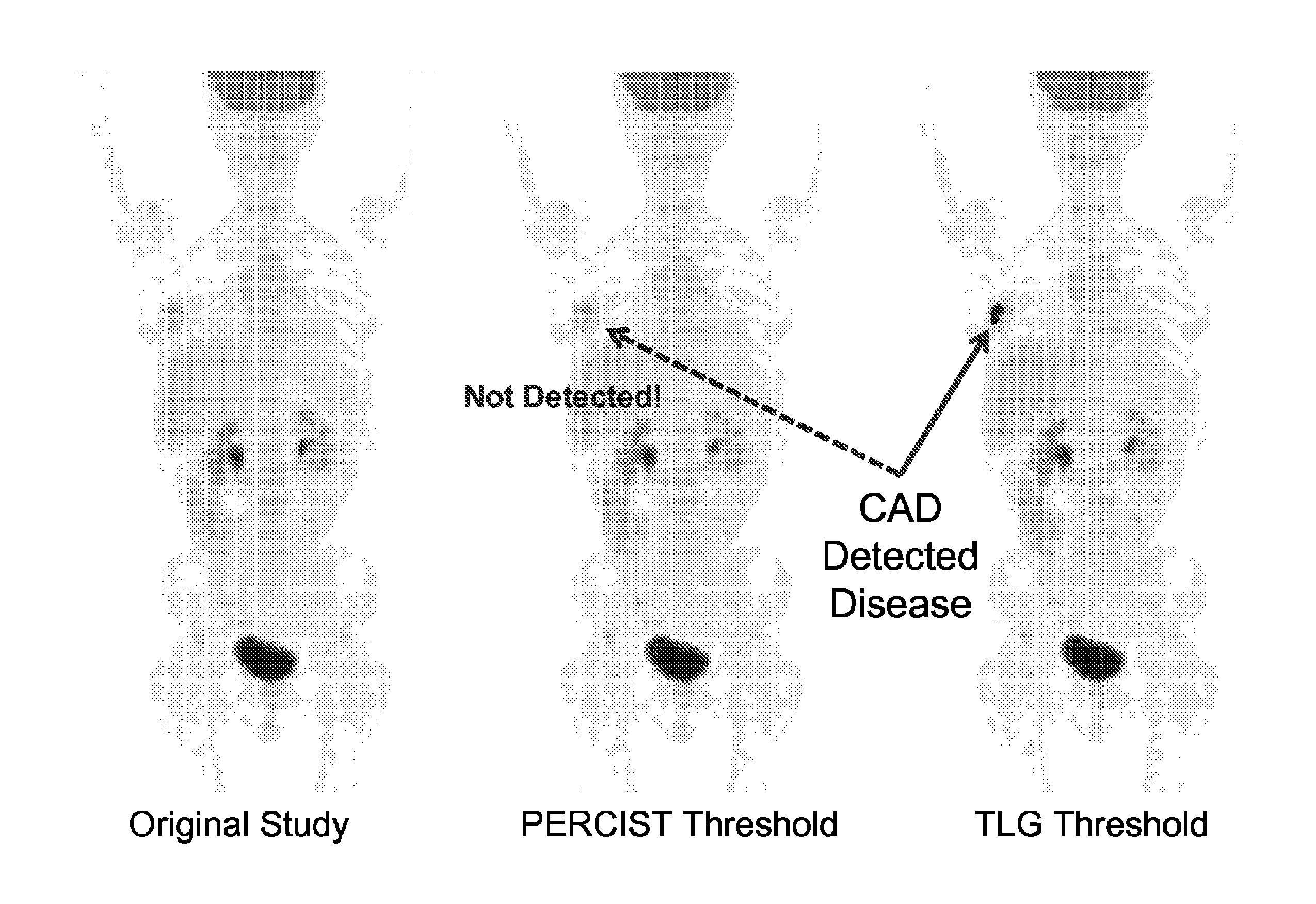
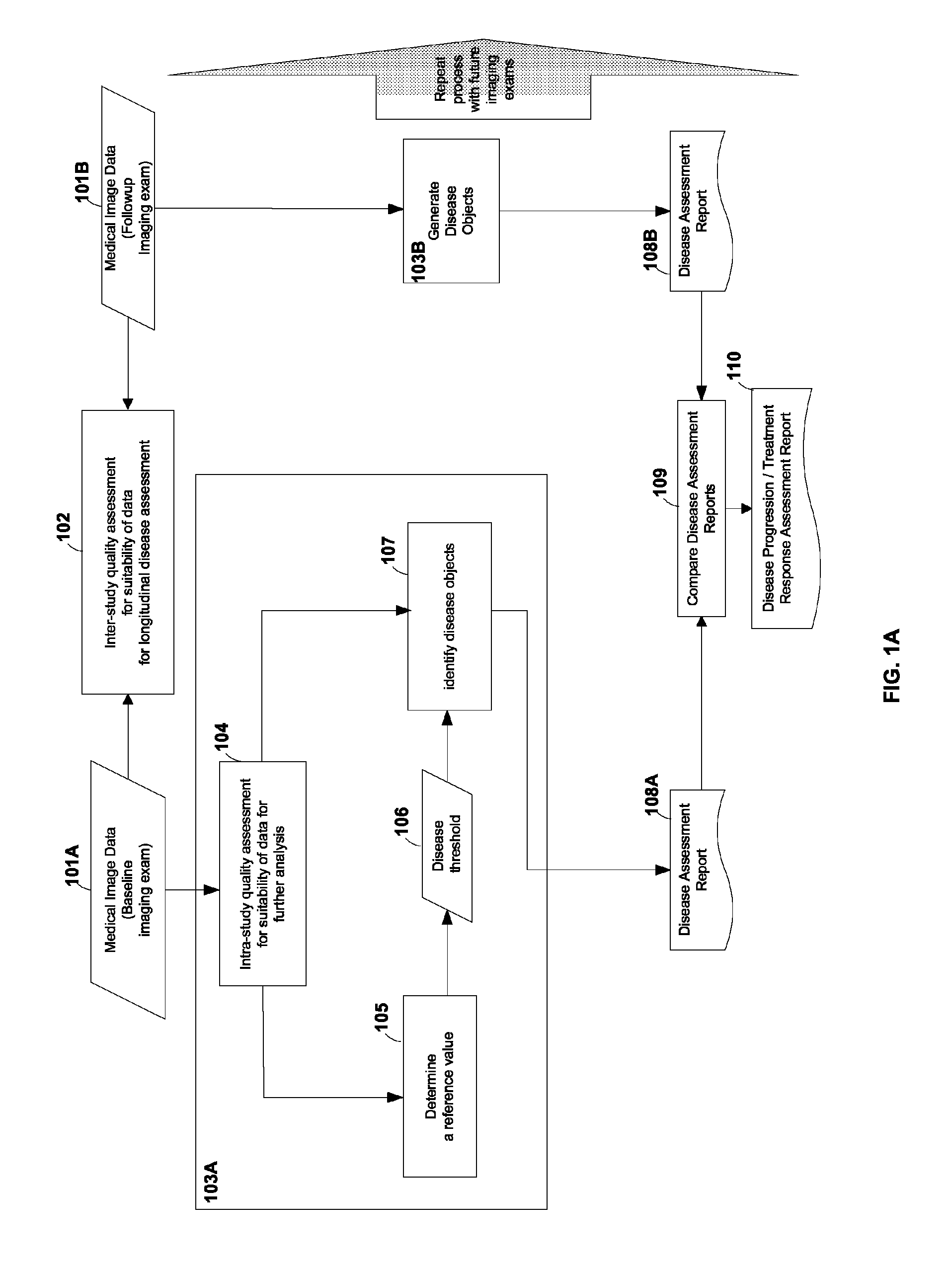
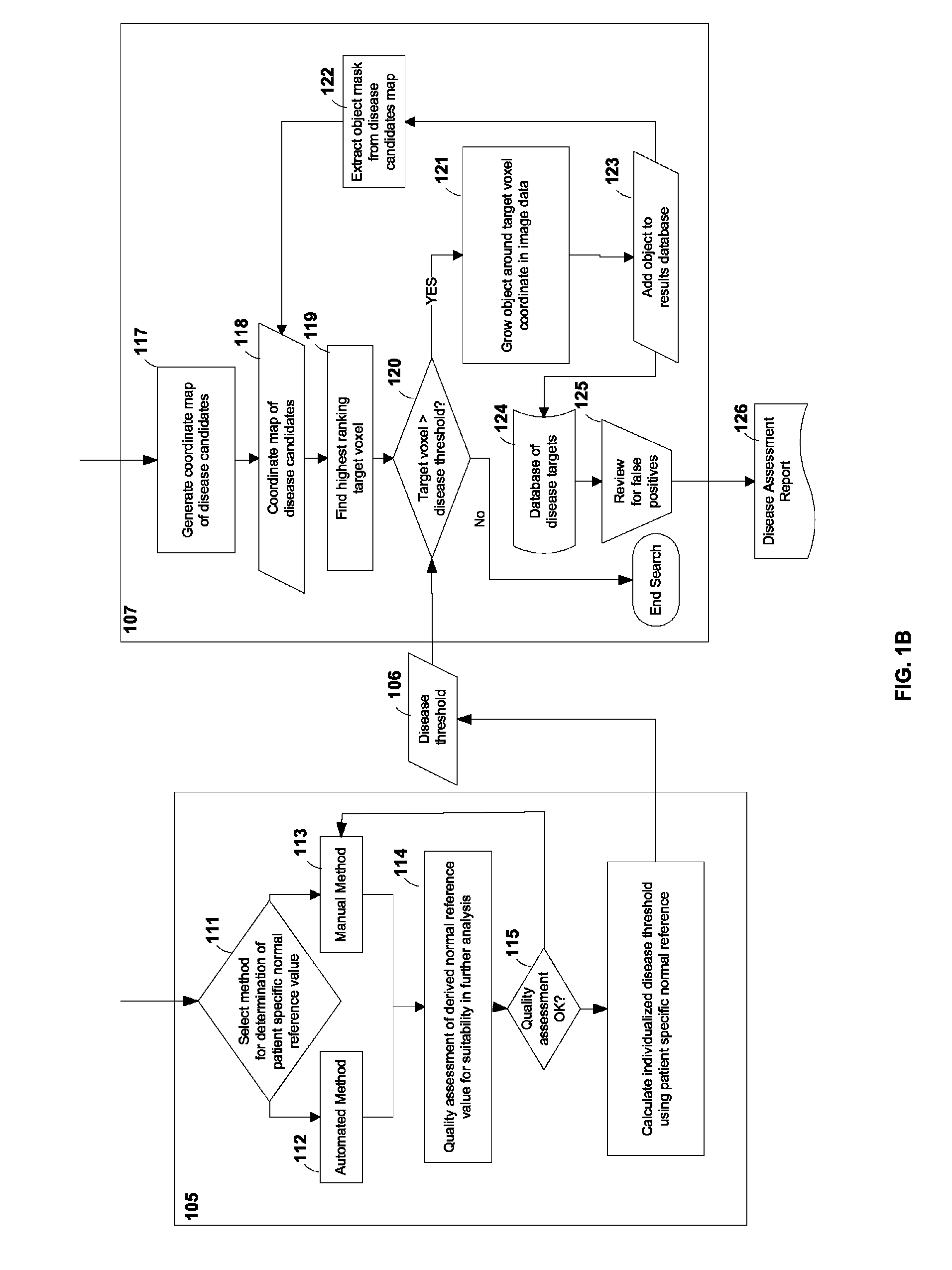
The current invention is a computer-implemented method for image processing, designed to detect and analyze disease tissue in medical images. The method involves receiving a first medical image, containing a large number of image voxels and representing different tissue regions of a person. The method automatically creates a reference value based on the first image, which defines a background level of intensity values for non-diseased tissue regions. It then generates a disease threshold based on the reference value. The method identifies specific areas of the image that correspond to disease tissue, which are made up of connected image voxels. The data about these disease tissue regions is then entered into a database for further comparison and analysis. Overall, the method helps to accurately detect and analyze abnormal tissue regions in medical images, which can aid in the diagnosis and treatment of various diseases.

Problems solved by technology

Such trials are expensive, not only in cost but in time.
Typically, the statistical power of phase I drug trials is inadequate to assess antitumor efficacy.
New drugs with a low response rate are typically not moved forward to advanced clinical testing under such a paradigm.
Not all phase III trials are successful, but all are costly.
Determining which innovative cancer therapeutics should be advanced to pivotal large phase III trials can be unacceptably delayed if survival is the sole endpoint for efficacy.
Survival trials can also be complicated by deaths due to nonmalignant causes, especially in older patients in whom comorbidities are common.
Additional complexities can include patients who progress on a clinical trial but who go on to have one of several nonrandomly distributed follow-up therapies—which can confound survival outcomes.

Method used


Image

  • Computer-aided detection (CAD) system for personalized disease detection, assessment, and tracking, in medical imaging based on user selectable criteria
  • Computer-aided detection (CAD) system for personalized disease detection, assessment, and tracking, in medical imaging based on user selectable criteria
  • Computer-aided detection (CAD) system for personalized disease detection, assessment, and tracking, in medical imaging based on user selectable criteria

Examples


Embodiment Construction

[0023]Some embodiments of the current invention are discussed in detail below. In describing the embodiments, specific terminology is employed for the sake of clarity. However, the invention is not intended to be limited to the specific terminology so selected. A person skilled in the relevant art will recognize that other equivalent components can be employed and other methods developed without departing from the broad concepts of the current invention. All references cited herein are incorporated by reference as if each had been individually incorporated.

[0024]FIG. 1A shows a flow-chart according to an embodiment of the current invention. Blocks 101A and 101B may represent medical image data over a time course of a subject under observation. The subject may be a human patient, an animal, etc. For example, block 101A may represent the medical image of a human patient with an abnormality before treatment. The medical image may comprise a plurality of image voxels and may represent m...


PUM


Abstract

An embodiment of the current invention includes computer-implemented method for image processing. The method includes receiving a first medical image from a data storage device, the first medical image comprising a plurality of image voxels and representing a plurality of tissue regions of a subject; automatically determining a reference value based on the first medical image, the reference value capable of providing a range of background level of voxel intensity values within at least one non-disease tissue region of the subject; generating a disease threshold based on the reference value; identifying portions of the medical image corresponding to disease-tissue regions according to the disease threshold, each of the portions comprising a plurality of connected image voxels in the medical image; and entering data encoding the disease-tissue regions into a database for subsequent comparisons.

Description

CROSS-REFERENCE OF RELATED APPLICATION[0001]This application claims priority to U.S. Provisional Application No. 61 / 406,693 filed on Oct. 26, 2010, and U.S. Provisional Application No. 61 / 453,647 filed on Mar. 17, 2011, the entire contents of both provisional applications are hereby incorporated by reference, and is a U.S. National Stage Application under 35 U.S.C. §371 of PCT / US2011 / 057890, filed Oct. 26, 2011, the entire contents of which are incorporated herein by reference.BACKGROUND[0002]1. Field of Invention[0003]The current patent application generally relates to medical image processing.[0004]2. Discussion of Related Art[0005]The ultimate goal of new cancer therapies is cure. A good cancer treatment should ideally prolong survival while preserving a high quality of life cost-effectively. To demonstrate prolonged survival in a clinical trial in some more slowly progressing cancers can take 5-10 years or longer. Such trials are expensive, not only in cost but in time.[0006]The...

Claims


Application Information

Patent Timeline
Patent Type & Authority Patents(United States)
IPC IPC(8): G06T7/00A61B6/00G06F19/00
CPCG06T7/0014A61B6/5217G06F19/3487A61B2576/00G06F19/321G06T2207/10072G06T2207/30016G06T2207/30096G16H15/00G16H50/20G16H30/40
Inventor LEAL, JEFFREYWAHL, RICHARD L.
Owner THE JOHN HOPKINS UNIV SCHOOL OF MEDICINE
Learn More