
Non-invasive optical imaging for measuring pulse and arterial elasticity in the brain

a technology of optical imaging and brain elasticity, applied in the field of systems, can solve the problems of limiting the use of continuous, non-invasive, portable, low-cost medical monitors, and inability to create accurate spatial maps, and fmri requires high-energy magnetic fields

Active Publication Date: 2015-10-27
THE BOARD OF TRUSTEES OF THE UNIV OF ILLINOIS
View PDF4 Cites 9 Cited by
  • Summary
  • Abstract
  • Description
  • Claims
  • Application Information

AI Technical Summary

Benefits of technology

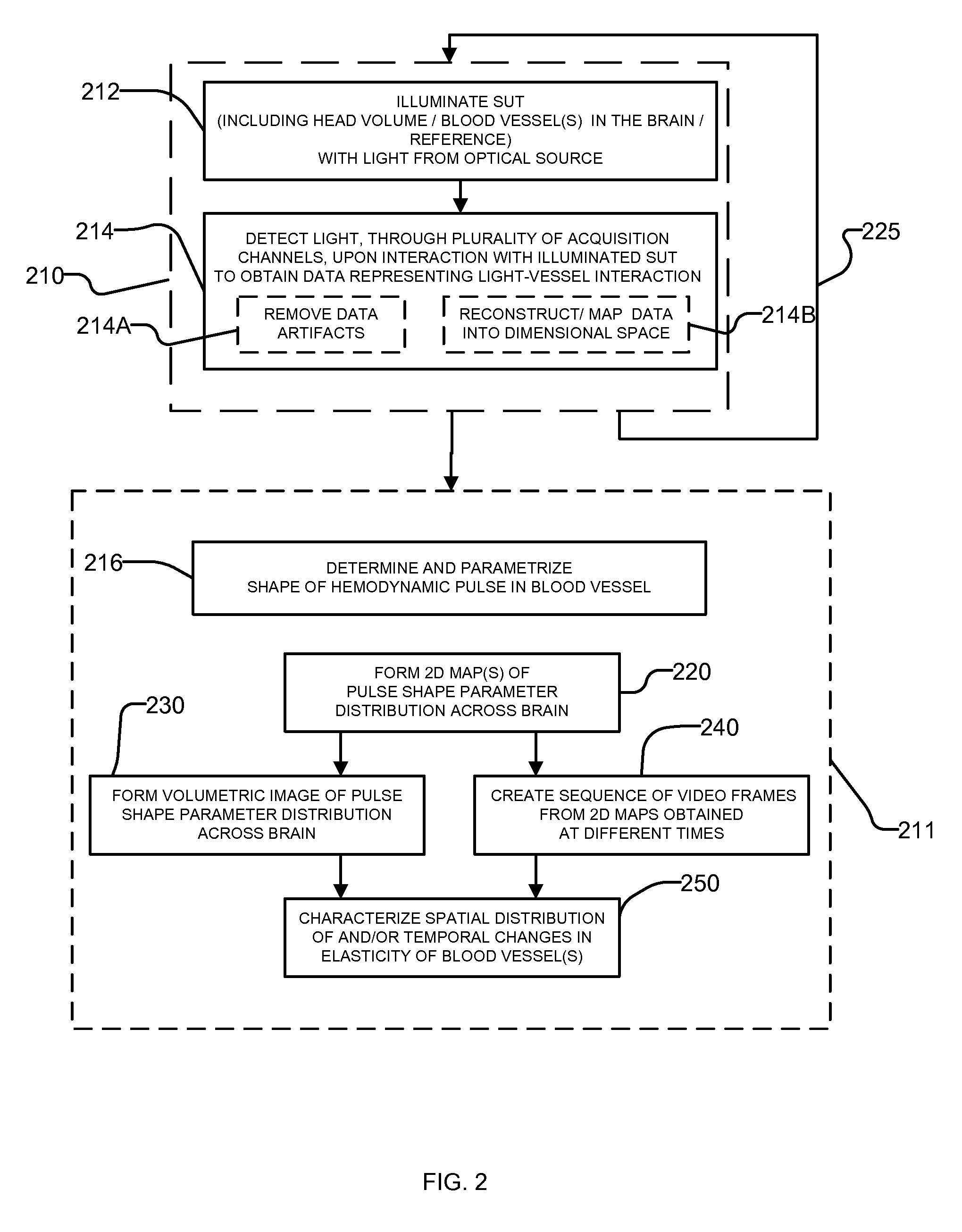
The invention provides a non-invasive method for measuring the elasticity of blood vessels in the brain using light imaging technology. By illuminating the brain with light and analyzing the resulting interaction data, the method can provide a valuable tool for studying brain blood flow and can help to diagnose and treat various brain disorders. The invention also provides an imaging system and computer program product for this purpose.

Problems solved by technology

All these techniques are powerful and have some advantages, but they suffer from drawbacks that limit their use as continuous, non-invasive, portable, and low-cost medical monitors.
For example, EEG can be inaccurate and does not lend itself to creating accurate spatial maps.
On the other hand, while PET and fMRI lend themselves to providing useful spatial maps of changes in brain functions, such as metabolism or blood flow, fMRI requires high-energy magnetic fields and PET data cannot be obtained quickly enough for informative comparison with other type of data.
Furthermore, PET requires the use of a radionuclide or radiopharmaceutical, which can be undesirable.
Likewise, CT requires the use of an ionizing radiation, which can also be undesirable.
However, ultrasound-based methods, such as TCD, produce only localized images and do not lend themselves to spatial mapping.
Furthermore, TCD is often disfavored in clinical settings due to the level of interference presented by the skull bones that significantly deform and disperse the propagation of the ultrasound waves thus making it complicated to provide localized estimates of intracranial structures.
Rather, when utilizing ultrasound to study arterial stiffness, the ultrasound analysis methods are typically limited to localized studies of arteries providing blood flow to the brain, such as arteries in the neck.

Method used


Image

  • Non-invasive optical imaging for measuring pulse and arterial elasticity in the brain
  • Non-invasive optical imaging for measuring pulse and arterial elasticity in the brain
  • Non-invasive optical imaging for measuring pulse and arterial elasticity in the brain

Examples


Embodiment Construction

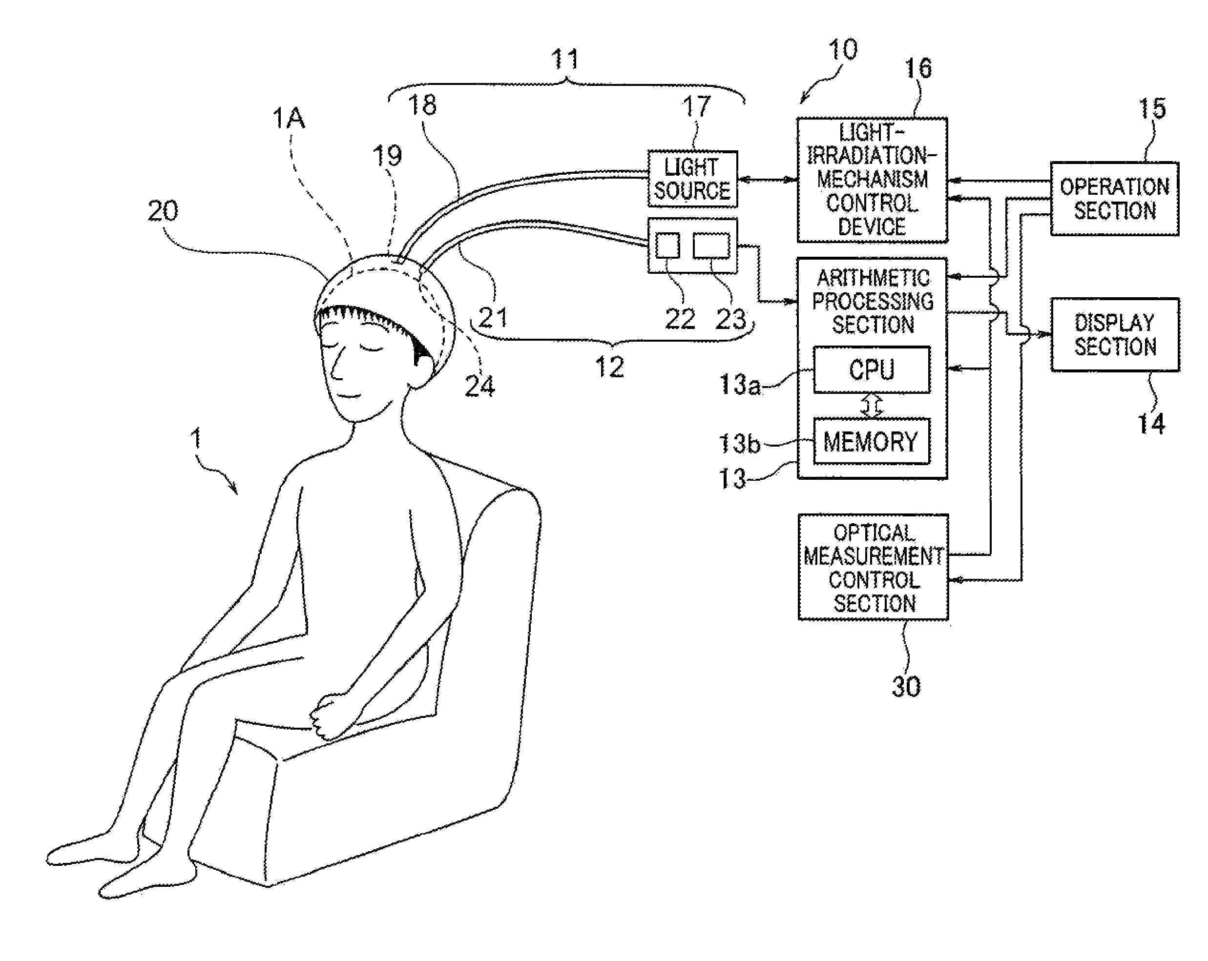
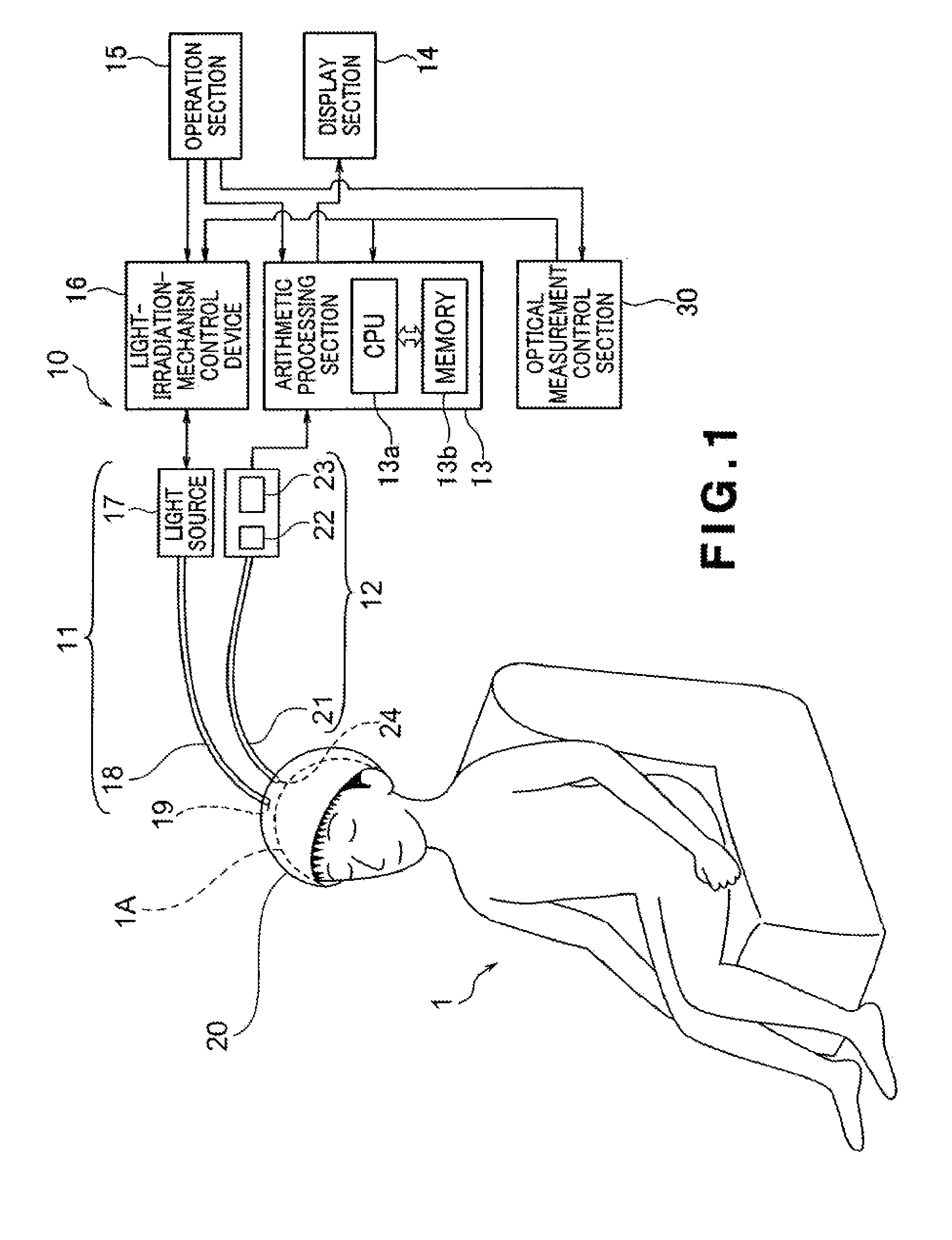
[0023]Recent advances in brain imaging studies suggest that understanding brain arterial stiffness or, alternatively, arterial elasticity can be a powerful indicator of cerebrovascular disease. The present invention provides a system and method for the incorporation of pulse data in brain mapping obtained with the use of diffusive optical methods to provide information about vascular health of different regions of the brain. Accordingly, a three-dimensional (3D) brain-mapping technique of the present invention makes use of optical tomography (OT) or diffuse OT (DOT) to acquire data associated with propagation of the pulse waveform through the brain.

[0024]Optical tomography and, in particular, DOT are centered around the idea that light passes through a body in small amounts and emerges bearing clues about tissues through which it has passed. These optical methods utilize non-ionizing NIR light, which is well tolerated in large doses by brain tissue, and have been recognized by some ...


PUM


Abstract

An optical imaging system and a method for generating a report regarding elasticity of arteries in the brain of a subject under test. Light output from the light source of the imaging system non-invasively illuminates at least one blood vessel or region of interest in the brain and, upon the interaction with the vessel, is registered with an optical detector to obtain a shape parameter of a hemodynamic pulse in the vessel. The shape parameter is further correlated to an elasticity parameter of the blood vessel(s) feeding the brain region of interest and presented in a form of report that may be a two- or a three-dimensional image of the parameter across the tested region of the brain.

Description

CROSS-REFERENCES TO RELATED APPLICATIONS[0001]This application represents the national stage entry of PCT International Application No. PCT / US2010 / 052750 filed on Oct. 14, 2010 and claims the benefit of U.S. Provisional Patent Application Ser. No. 61 / 256,024 filed on Oct. 29, 2009. The above-referenced patent applications are incorporated herein by reference in their entirety.STATEMENT REGARDING FEDERALLY SPONSORED RESEARCH OR DEVELOPMENT[0002]This invention was made with support under The National Institutes of Health (NIH) grant AG021887. The United States Government has certain rights in the invention.TECHNICAL FIELD[0003]The present invention relates to systems and methods for performing studies of brain tissue and, in particular, to optical systems and methods for measuring elasticity of arteries in the brain.BACKGROUND ART[0004]Normal aging is accompanied by a number of cognitive changes, including decline in working memory and executive function / attention control, as well as ...

Claims


Application Information

Patent Timeline
Patent Type & Authority Patents(United States)
IPC IPC(8): A61B5/00A61B5/02
CPCA61B5/0059A61B5/02007A61B5/02028A61B5/7207A61B5/0042A61B5/4064
Inventor GRATTON, GABRIELEFABIANI, MONICALOW, KATHYMACLIN, EDWARD
Owner THE BOARD OF TRUSTEES OF THE UNIV OF ILLINOIS
Learn More