Tumor antigens elicit anti-tumor humoral immune reactions in a subset of patients with polycythemia vera

a technology of humoral immune reaction and tumor antigen, which is applied in the field of self-tumor antigens, can solve the problems of limited use practicability of internal ribosome entry sites, and achieve the effect of higher expression and higher expression

Active Publication Date: 2015-10-20
TEMPLE UNIVERSITY
View PDF2 Cites 0 Cited by
  • Summary
  • Abstract
  • Description
  • Claims
  • Application Information

AI Technical Summary

Benefits of technology

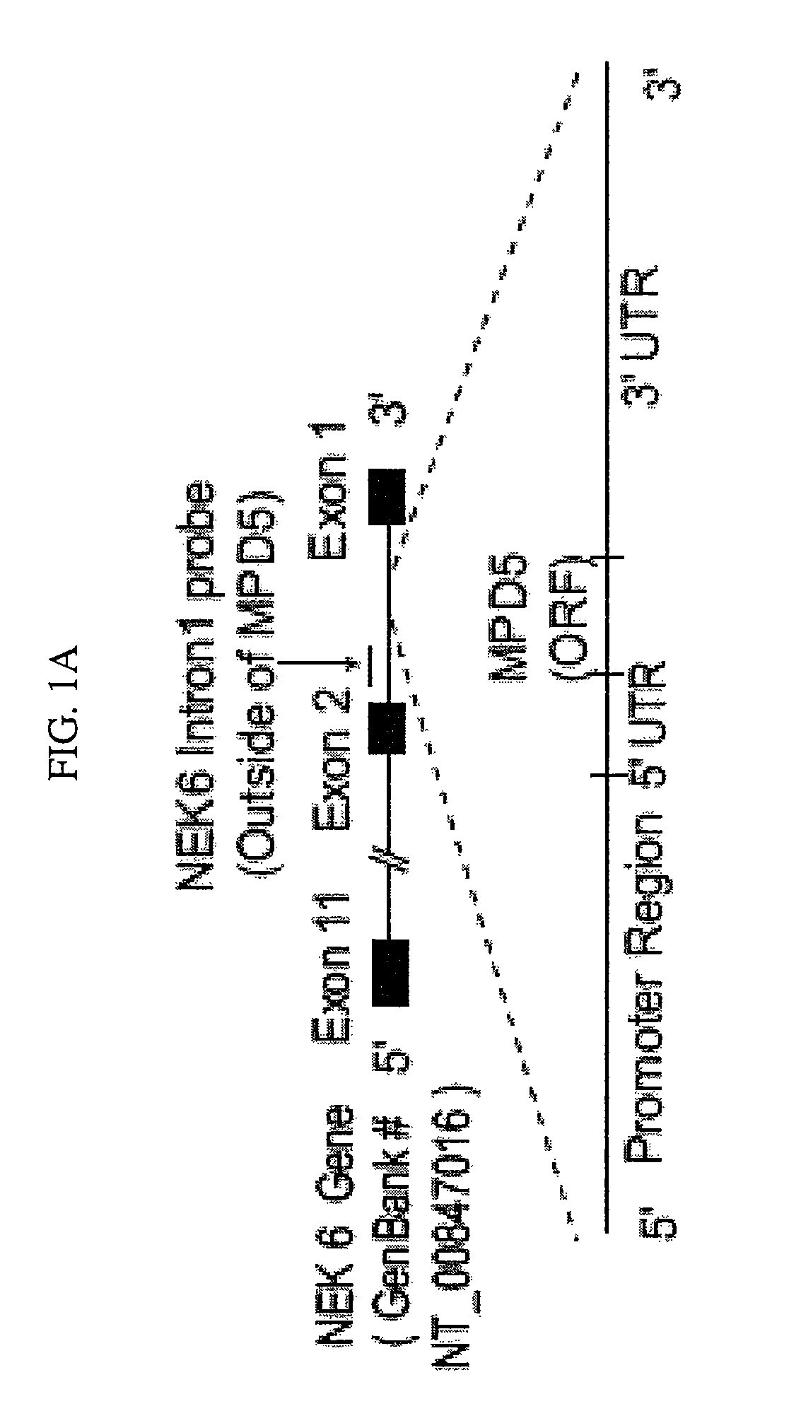
[0039]FIG. 2. The expression of MPD5 transcripts is elevated in tumors and PV. FIG. 2A. The expression of MPD5 transcripts in normal tissues. Lanes N1 to N10 indicate various normal tissues in the brain (N1), liver (N2), placenta (N3), small intestine (N4), colon (N5), thymus (N6), spleen (N7), prostate (N8), testis (N9), and ovary (N10), respectively. The transcript sizes are indicated as kilobases (kb). FIG. 2B. Expression of MPD5 transcripts in tumor cells, as detected by Northern blot. Lanes T1 to T10 indicate various tumor cells in acute T cell leukemia (Jurkat cells) (T1), Burkitt's lymphoma (CA46) (T2), breast cancer (MDA-MD-453) (T3), Burkitt's lymphoma (Namalwa) (T4), epidermal carcinoma (A-431) (T5), uterine carcinoma (MES-SA) (T6), Burkitt's lymphoma (Raji) (T7), osteosarcoma (MG-63) (T8), histiocytic lymphoma (U-937) (T9), and cervical adenocarcinoma (Hela S3) (T10), respectively. In the lower panel, the specificity of the NEK6 intron 1 probe (outside of MPD5) was confirmed by Southern blot of an agarose gel analyzing a NEK6 intron 1 fragment that was located outside the MPD5 region and had no overlap with MPD5. FIG. 2C. The expression of MPD5 transcripts in the granulocytes from PV patients and healthy donors, as detected by quantitative RT-PCR. The expression levels of MPD5 transcripts are expressed as the ΔCT (MPD5-18S). Low ΔCT values indicate higher expression of the specific gene. The groups, whose expression of MPD5 transcripts is statistically different from that of healthy donors (p<0.05), are marked with an *. FIG. 2D. The MPD5 expression in K562 cells stimulated with IFN-α, as detected by semi-quantitative RT-PCR (the left panel). RT-PCR of β-actin served as a housekeeping control for IFN-α stimulation. The RT-PCR of ISG15 was used as a positive control for IFN-α stimulation. In the right panel, the densitometric units were calculated by normalizing the densities of the PCR products of MPD5 and ISG15 with those of β-actin in the same sample.
[0040]FIG. 3. MPD5 peptide specifically reacts to sera from PV patients responding to therapies. IgG antibody responses to the C-terminal antigenic epitope (from aa 30 to aa 47) of MPD5 were detected by peptide ELISA. These experiments were repeated three times, and representative results are shown. The mean plus three standard deviations (SD) of the OD405 peptide ratios over the coating control (derived from 23 healthy donors) was calculated as the upper limit of the normal range of MPD5 peptide antibody responses (the mean+3SD=1.38). The groups with detection rates of IgG antibody responses to MPD5 peptide that are statistically higher than those of healthy donors (the chi-square goodness-of-fit test; p<0.05) are marked with an asterisk *.
[0041]FIG. 4. The novel tumor antigen PV65 (eIF-2α). FIG. 4A. Schematic representation of the genomic structure, mRNA, and protein structure of tumor antigen PV65 (eIF-2α, GenBank accession number: NM—032025). FIG. 4B. The expression of PV65 transcripts in normal tissues detected by Northern blot. The lanes N1 to N10 indicate various normal tissues in the order of brain (N1), liver (N2), placenta (N3), small intestine (N4), colon (N5), thymus (N6), spleen (N7), prostate (N8), testis (N9), and ovary (N10), respectively. The hybridization analyses of the normal tissue and tumor cell expression (BD Clontech) with 32P-Labelled specific probes, as indicated, were performed, respectively. The transcript sizes are indicated with kilobases (kb). FIG. 4C. The expression of PV65 transcripts in tumor cells detected by Northern blot. The lanes T1 to T10 indicate various tumor cells in the order of acute T cell leukemia (Jurkat cells) (T1), Burkitt's lymphoma (CA46) (TI), breast cancer (MDA-MD-453) (T3), Burkitt's lymphoma (Namalwa) (T4), epidermal carcinoma (A-431) (T5), uterine carcinoma (MES-SA) (T6), Burkitt's lymphoma (Raji) (T7), osteosarcoma (MG-63) (T8), histiocytic lymphoma (U-937) (T9), and cervical adenocarcinoma (Hela S3) (T10), respectively. FIG. 4D. The expression of PV65 transcripts in the granulocytes from PV patients and healthy donors detected by quantitative RT-PCR. The expression levels of PV65 transcripts are expressed as the deltaCT (PV65-18S). Low deltaCT values indicate higher expression of the specific gene. The experiments were repeated for three times. The mean and standard deviation for each group were calculated. FIG. 4E. Western blot analyses of PV65 protein and phosphorylated PV65 protein in K562 myeloid leukemia cells. The expression levels of PV65 protein and phosphorylated PV65 protein in K562 cells at 0, 2, 4, and 8 hours after stimulation by IFN-α were assayed with Western blots using anti-PV65 and antiphosphorylated PV65, respectively. The Western blot analysis for the house keeping protein control β-actin was also performed using anti-β-actin as protein loading control. FIG. 4F. The IgG antibody reactions to the tumor antigen PV65 detected by phage plaque assay. The detection rates in each group are presented with the empty column as the positive (on the top) and the solid column as the negative (on the bottom). The experiments were repeated for three times. The representative results are shown. The groups, whose detection rates of the IgG antibody reactions to PV65 are statistically higher than that of healthy donors (the Chi-Square Goodness of-Fit Test; p<0.05), are marked with *.
[0042]FIG. 5. The novel tumor antigen PV13 (protamine 2). FIG. 5A. Schematic representation of the genomic structure, mRNA, and protein structure of tumor antigen PV13 (protamine 2, GenBank accession number: NM—002762). FIG. 5B. The expression of PV13 transcripts in normal tissues detected by Northern blot. The lanes N1 to N10 indicate various normal tissues in the order of brain (N1), liver (N2), placenta (N3), small intestine (N4), colon (N5), thymus (N6), spleen (N7), prostate (N8), testis (N9), and ovary (N10), respectively. The hybridization analyses of the normal tissue and tumor cell expression (BD Clontech) with 32P-labelled specific probes, as indicated, were performed, respectively. The transcript sizes are indicated with kilobases (kb). FIG. 5C. The expression of PV13 transcripts in tumor cells detected by Northern blot. The lanes T1 to T10 indicate various tumor cells in the order of acute T cell leukemia (Jurkat cells) (T1), Burkitt's lymphoma (CA46) (T2), breast cancer (MDA-MD-453) (T3), Burkitt's lymphoma (Namalwa) (T4), epidermal carcinoma (A-431) (T5), uterine carcinoma (MES-SA) (T6), Burkitt's lymphoma (Raji) (TI), osteosarcoma (MG-63) (T8), histiocytic lymphoma (U-937) (T9), and cervical adenocarcinoma (Hela S3) (T10), respectively. FIG. 5D. The IgG antibody reactions to the tumor antigen PV13 detected by phage plaque assay. The detection rates in each group are presented with the empty column as the positive (on the top) and the solid column as the negative (on the bottom). The experiments were repeated for three times. The representative results are shown. The groups, whose detection rates of the IgG antibody reactions to PV13 are statistically higher than that of healthy donors (the Chi-Square Goodness-of-Fit Test; p<0.05), are marked with *.

Problems solved by technology

The practical use of internal ribosome entry site is limited in construction of bicistronic vectors for gene therapy and construction of bicistronic vectors for gene expression.

Method used


Image

  • Tumor antigens elicit anti-tumor humoral immune reactions in a subset of patients with polycythemia vera
  • Tumor antigens elicit anti-tumor humoral immune reactions in a subset of patients with polycythemia vera
  • Tumor antigens elicit anti-tumor humoral immune reactions in a subset of patients with polycythemia vera

Examples


examples

Serum Samples

[0130]In accordance with a protocol approved by the Institutional Review Board at Temple University and established guidelines, serum samples were obtained from patients with PV and CML receiving IFN-α therapy enrolled into Baylor College of Medicine (eight patients with PV receiving IFN-α treatment, 8 PV patients receiving other treatments), A. D. Anderson Cancer Center (! 0 patients with C!\L receiving TFN-(X treatment), New York Presbyterian Hospital-Weill Cornell Medical Center (13 PV patients receiving IFN-α treatment, 26 PV patients receiving other treatments, six PV patients having not yet received any treatments) Institutional Review Board-approved trials. The therapeutic regimens other than IFN-α for the 34 patients with PV included imatinib mesylate (Gleevec, 16 patients), hydroxyurea ci 2 patients), hydroxyurea plus agrelin (three patients), agrelin plus phlebotomy (one patient), phlebotomy (one patient), and thalidomide (one patient). The patients in the oth...


PUM

PropertyMeasurementUnit
temperaturesaaaaaaaaaa
diameteraaaaaaaaaa
fluorescentaaaaaaaaaa

Abstract

The invention provides novel antigens, MPD5, PV13, and PV65, which belongs to the group of cryptic antigens without conventional genomic structure and is encoded by a cryptic open reading frame located in the 3′untranslated region (3′UTR) of myotrophin mRNA. The antigens elicit IgG antibody responses in a subset of PV patients, as well as patients with chronic myelogenous leukemia and prostate cancer. The translation of MPD5, PV13 and PV65 was mediated by a novel internal ribosome entry site (IRES) upstream of the open reading frame. Eliciting anti-tumor immune response against MPD5, PV13 and/or PV65 antigen in patients with myeloproliferative diseases is a novel immunotherapy.

Description

CROSS-REFERENCE TO RELATED APPLICATIONS[0001]This application claims benefit of U.S. Provisional Patent Application No. 60 / 863,194 filed Oct. 27, 2006, and U.S. Provisional Patent Application No. 60 / 868,585 filed Dec. 5, 2006, the entire disclosures of which are hereby incorporated by reference in their entireties.STATEMENT REGARDING FEDERALLY SPONSORED RESEARCH OR DEVELOPMENT[0002]This research was supported in part by U.S. Government funds (NIH Grant No. AI054514), and the U.S. Government may therefore have certain rights in the invention.BACKGROUND OF THE INVENTION[0003]1. Field of Invention[0004]This invention relates to self-tumor antigens and methods and compositions to generate immunity in humans against self tumor antigens. This invention is more particularly related to eliciting or enhancing immunity against human self tumor antigen encoded by unconventional reading frames with homology to foreign proteins[0005]2. Description of Related Art[0006]Self-tumor antigens that eli...

Claims


Application Information

Patent Timeline
Patent Type & Authority Patents(United States)
IPC IPC(8): C07K7/00C07K16/30
CPCC07K16/30A61P37/04
Inventor YANG, XIAO-FENG
Owner TEMPLE UNIVERSITY
Why Eureka
Try Eureka