Indices for management of dry eye syndrome, corneal ectasia, keratoplasty graft rejection and failure and Fuchs' dystrophy
a technology of keratoplasty graft and index, which is applied in the field of index for management of dry eye syndrome, corneal ectasia, keratoplasty graft rejection and failure, and fuchs' dystrophy, can solve the problems of adversely affecting the quality of life of patients, body sensation, and difficult diagnosis and treatment, and achieve improved indices, improved diagnosis, treatment and monitoring, and enhanced epithelial irregularity factor
- Summary
- Abstract
- Description
- Claims
- Application Information
AI Technical Summary
Problems solved by technology
Method used
Image

Examples
Embodiment Construction
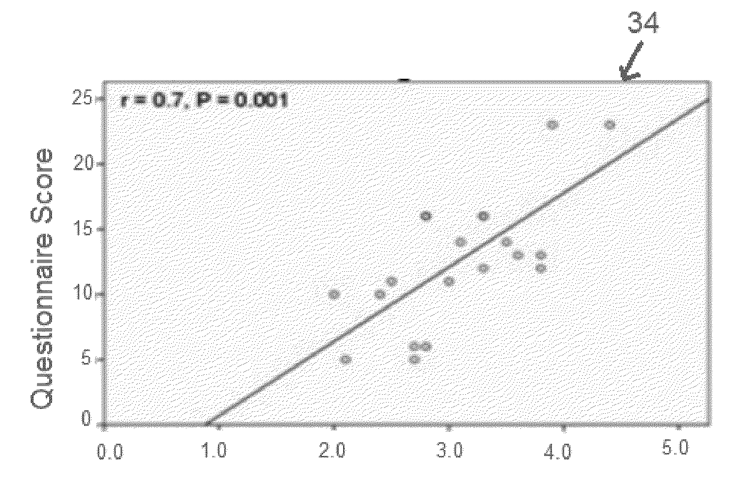
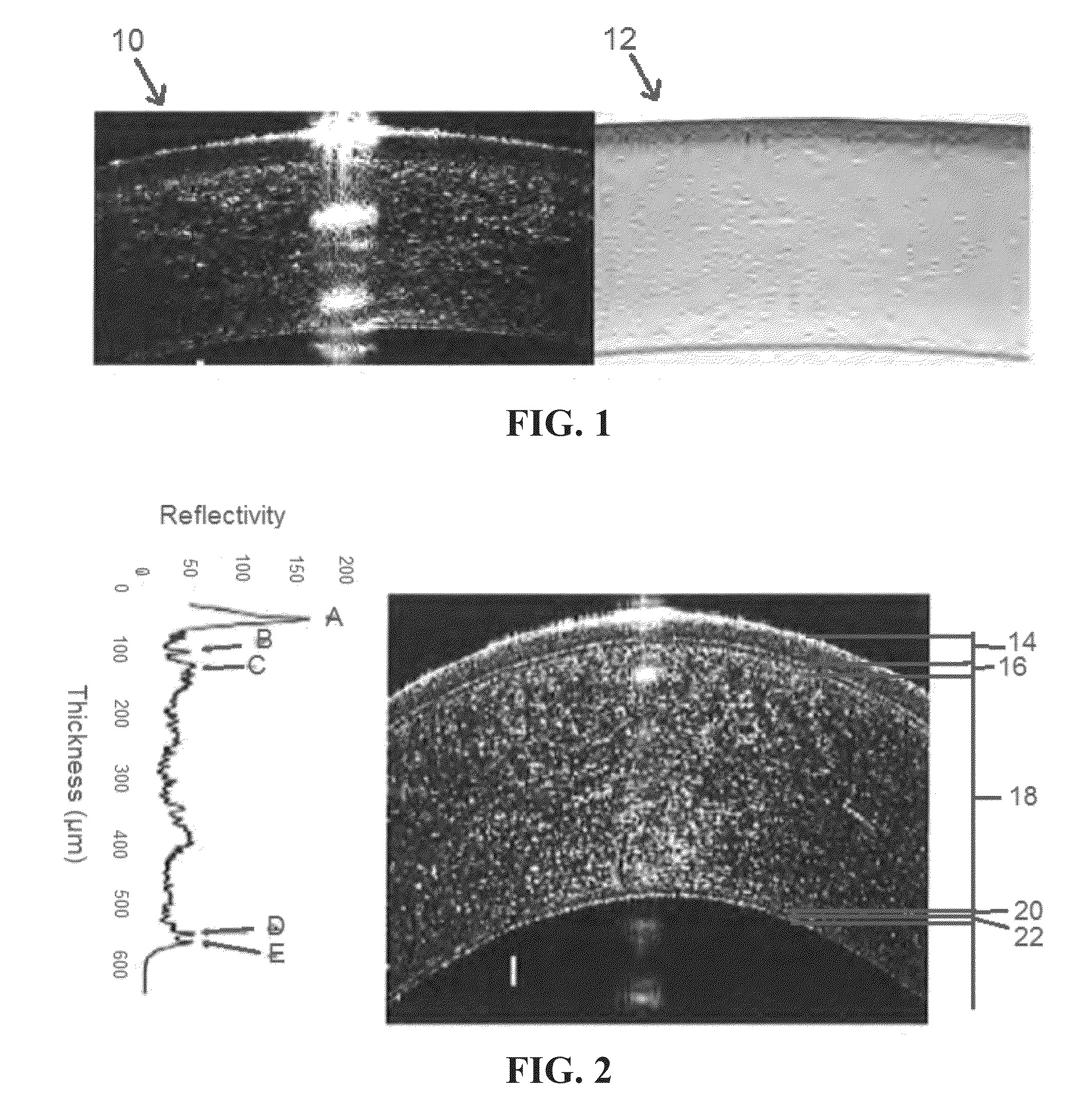
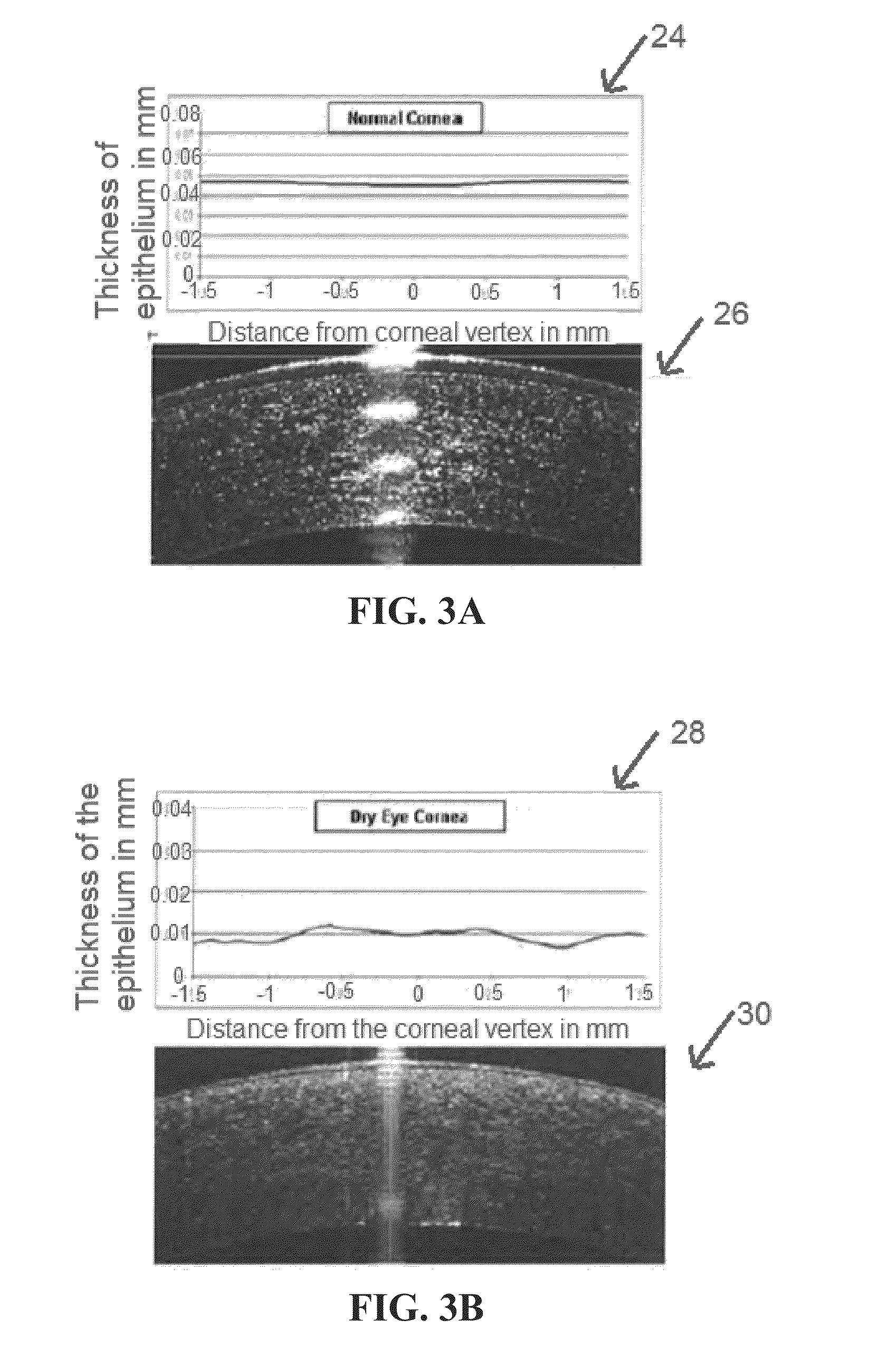
Enhanced Epithelial Irregularity Factor (eEIF)
[0038]Referring generally to FIGS. 1-6, figures relating to Enhanced Epithelial Irregularity Factor (eEIF) are shown. One aspect of the present invention is based on the observation that the corneal epithelium of patients with dry eye syndrome (DES) is irregular, and that irregularity can be quantified using an epithelial irregularity factor (EIF) in a standardized and objective method that correlates to the subjective symptoms of a patient. The corneal epithelium irregularity can be reversed by treatment, and therefore this factor can be used to monitor response to treatment and can be used to test new therapies.
[0039]In order to examine the epithelium, or other parts of the eye, an imaging system such as an optical coherence tomography (OCT) system may be used, including ultrahigh-resolution OCT (UHR-OCT) systems. Additionally or alternatively, any other imaging device or system may be used that is capable of providing high-resolution ...
PUM
 Abstract
Description
Claims
Application Information
 - R&D
- Intellectual Property
- Life Sciences
- Materials
- Tech Scout
- Unparalleled Data Quality
- Higher Quality Content
- 60% Fewer Hallucinations
Browse by: Latest US Patents, China's latest patents, Technical Efficacy Thesaurus, Application Domain, Technology Topic, Popular Technical Reports.
© 2025 PatSnap. All rights reserved.Legal|Privacy policy|Modern Slavery Act Transparency Statement|Sitemap|About US| Contact US: help@patsnap.com