Method and apparatus for diagnosis of tumor activity using tumor interstitial fluid pressure

a technology of interstitial fluid and tumor activity, which is applied in the field of method and apparatus for diagnosis of tumor activity using tumor interstitial fluid pressure, can solve the problems of loss of oncotic pressure gradient across the blood vessel wall, abnormal tumor vasculature, and impaired lymphatic drainag

Inactive Publication Date: 2015-03-24
HENRY FORD HOSPITAL +1
View PDF1 Cites 0 Cited by
  • Summary
  • Abstract
  • Description
  • Claims
  • Application Information

AI Technical Summary

Benefits of technology

[0008]It has been recognized that after the initial filling or exposure of a tumor with a suitable contrast agent, as a result of the leaky vasculature characteristics of selected tumor blood vessels, the size of the detected or imaged region of contrast will increase with time. In particular, the inventors have appreciated that due to fluid flux from the tumor into the patient's surrounding normal tissues, following the initial concentration and scanning of the contrast agent, the detected imaged area of the tumor will over time expand in a halo effect. The present invention provides in one possible embodiment, an apparatus and method for measuring the rate and / or extent of observed contrast-enhanced volume expansion and / or intensity change of the dyed tissue area as an indication of one or more of tumor type, tumor activity, treatment effectiveness and / or treatment response.

Problems solved by technology

Blood proteins permeate leaky vasculature, resulting in a loss of oncotic pressure gradient across the blood vessel wall.
High TIFP impairs lymphatic drainage and may contribute to tumor vasculature being abnormal.
As tumors grow, the abnormal vasculature limits the delivery of metabolites (nutrition and oxygen supply) and removal of wastes (such as lactic acid causing tumors to become acidic) to the tumor center.

Method used


Image

  • Method and apparatus for diagnosis of tumor activity using tumor interstitial fluid pressure
  • Method and apparatus for diagnosis of tumor activity using tumor interstitial fluid pressure
  • Method and apparatus for diagnosis of tumor activity using tumor interstitial fluid pressure

Examples


Embodiment Construction

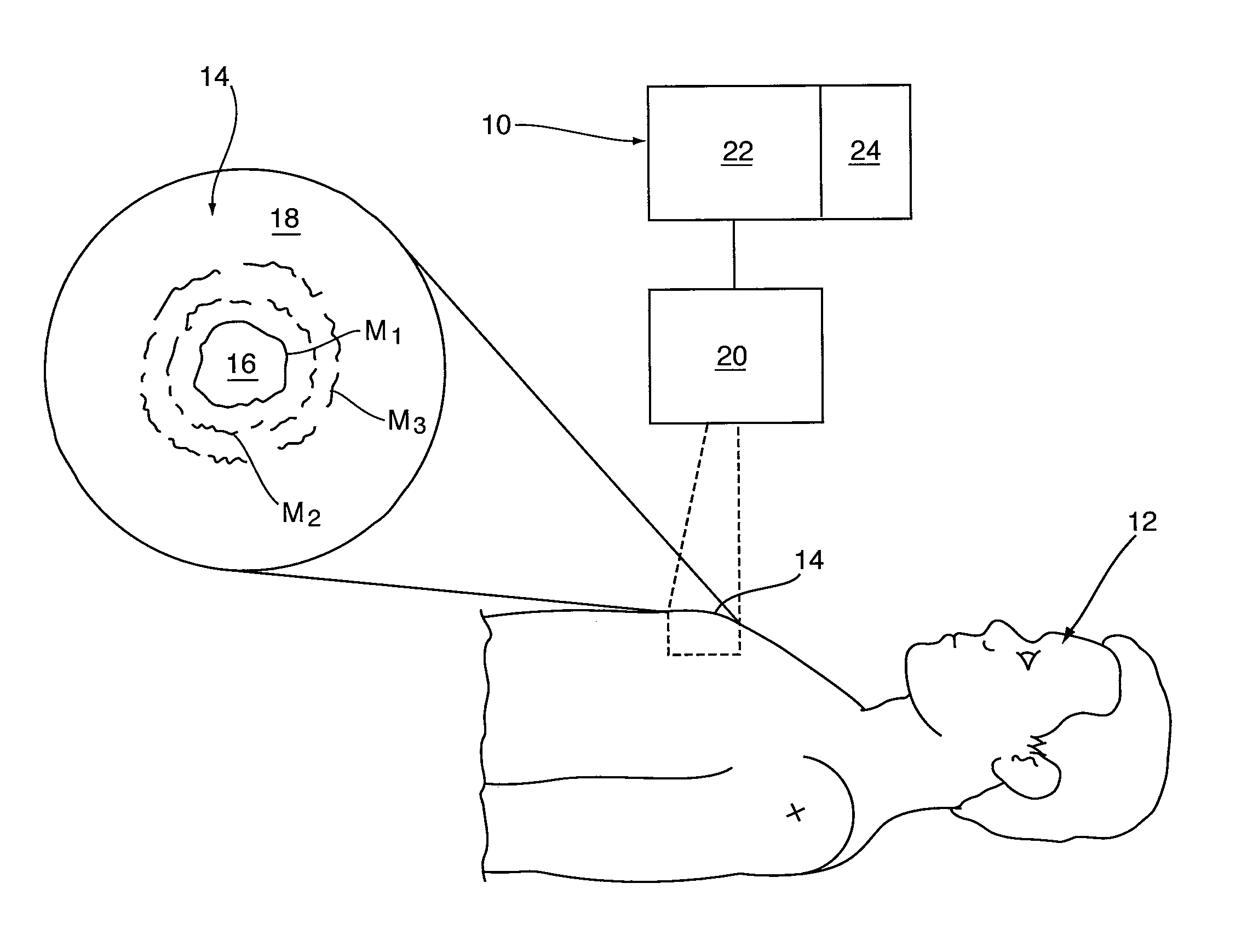
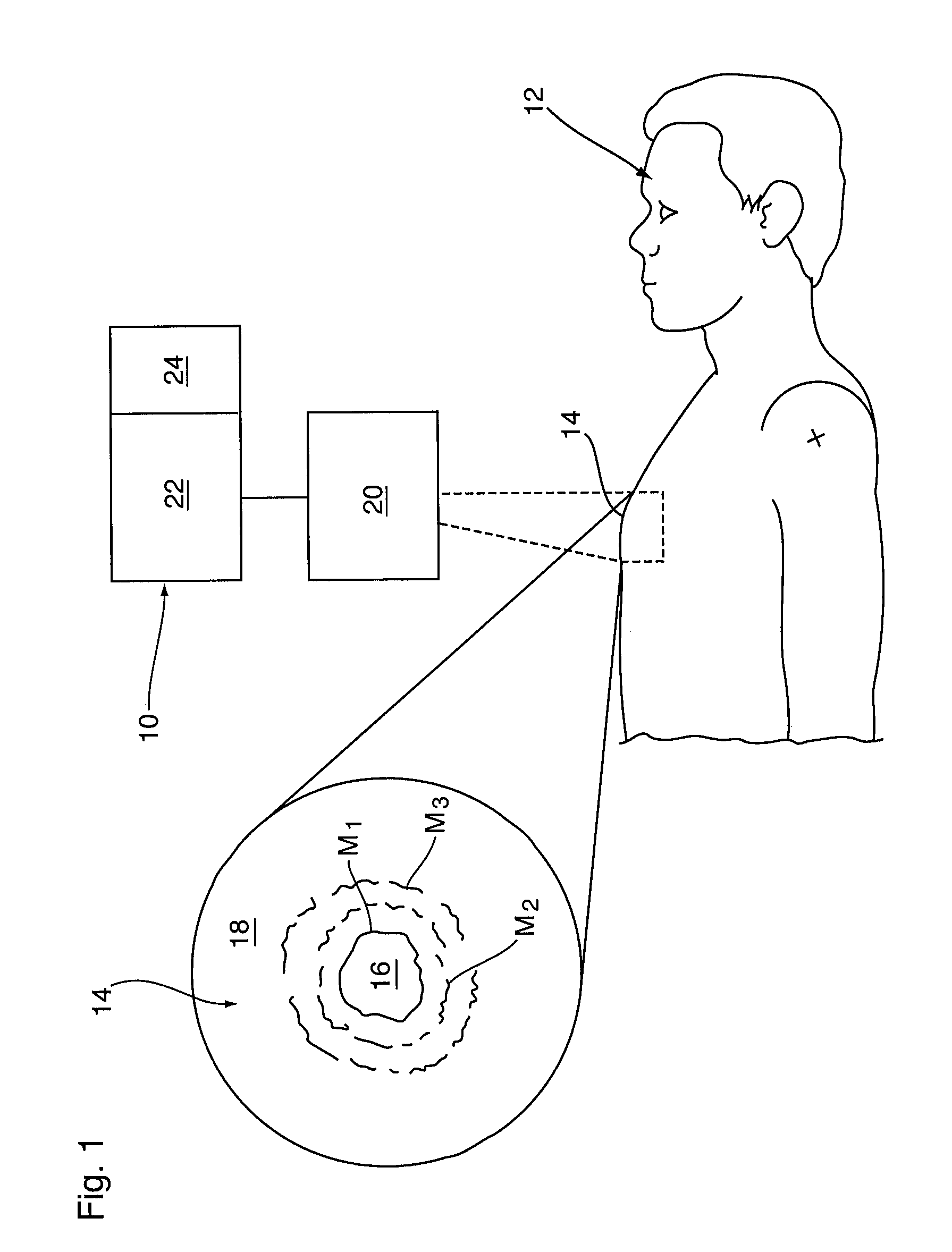
[0018]Reference may be had to FIG. 1 which illustrates schematically an apparatus 10 for use in tumor identification and / or diagnosis in a patient 12. As will be described, the apparatus 10 is used in conjunction with a suitable contrast agent which is selected both for injection into the patient 12 to initially collect and concentrate within tumor or cancerous tissues (hereinafter collectively a tumor 16), and to be detectable by the apparatus 10 as a tumor marker to provide a detectable or imaged marker M thereof. In a preferred embodiment, the apparatus 10 is provided with an electromagnetic radiation (EMR) source 20 which is operable to produce optical images of a region of interest 14 in the patient 12, and which is selected as a site likely including a solid tumor 16 together with its surrounding tissue 18.

[0019]The EMR source 20 is preferably selected as an MRI apparatus which is operable to detect and output an image or data (hereinafter collectively referred to as an image)...


PUM


Abstract

A scanning apparatus, is used to effect multiple images of a tumor in which a contrast agent has been localized as a detectable marker over a selected time to map the change in the imaged marker. The rate of change in the imaged marker and / or contrast intensity of the dyed tissues is used to assess tumor aggressiveness and as an early predictor of response to cancer therapy. In particular, following the marking of tumor or cancerous tissues by the initial localization of an imageable contrast agent, the rate of change in the volume and / or area of the imaged marker is used to provide an indication of tumor interstitial fluid pressure (TIFP).

Description

SCOPE OF THE INVENTION[0001]The present invention provides for the simplified measurement of the rate of fluid flux away from tumor tissues as an indication of tumor type, aggressiveness and / or treatment effectiveness, and more preferably as indicating relative differences between tumor interstitial fluid pressure and that of normal tissues.BACKGROUND OF THE INVENTION[0002]Despite remarkable strides in the treatment of solid cancers, such as those in the breast and prostate, many cancers remain resistant to treatment. As new therapies are developed and medicines become increasingly individualized, a need exists for effective and early diagnosis and / or predictor of treatment effectiveness or treatment response.[0003]One physiological parameter which has demonstrated predictive value for tumor type or aggressiveness, and response to chemotherapy and / or radiotherapy is tumor interstitial fluid pressure (TIFP). Tumor interstitial fluid pressure (TIFP) is a physiological parameter that i...

Claims


Application Information

Patent Timeline
Patent Type & Authority Patents(United States)
IPC IPC(8): A61B5/03A61B5/055A61B5/00
CPCA61B5/03A61B5/0059
Inventor SCHLESINGER, MORDECHAYLIU, LONG JIANEWING, JAMES R.BROWN, STEPHEN L.
Owner HENRY FORD HOSPITAL
Why Eureka
Try Eureka