
Methodology to identify the scleral spur

a scleral spur and scleral spur technology, applied in the field of ophthalmic diagnosis and treatment, can solve the problems of difficult scleral spur locating, inability to effectively image deeper ocular structures, and requiring significant training and experience, and achieve the effect of improving the examination of the ey

Active Publication Date: 2015-03-03
ESI
View PDF3 Cites 10 Cited by
  • Summary
  • Abstract
  • Description
  • Claims
  • Application Information

AI Technical Summary

Benefits of technology

The present invention improves ultrasonic biomicroscopy to better locate the scleral spur and aid in the diagnosis and treatment options of ocular disease. It also facilitates the location of the scleral-uveal interface, corneal curvature, and intersection point. The invention serves as an improved examination tool for the eye.

Problems solved by technology

OCT is a newer technology which is promising, but cannot effectively image deeper ocular structures.
UBM allows visualization behind the iris and is useful, but locating the scleral spur is difficult, requiring significant training and experience.
Accurately locating the subtle scleral spur structure with even the highest frequency ultrasound equipment is often difficult and as a consequence the location of the spur must often be estimated.

Method used


Image

  • Methodology to identify the scleral spur
  • Methodology to identify the scleral spur
  • Methodology to identify the scleral spur

Examples


Embodiment Construction

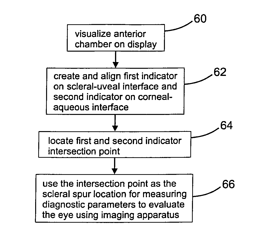
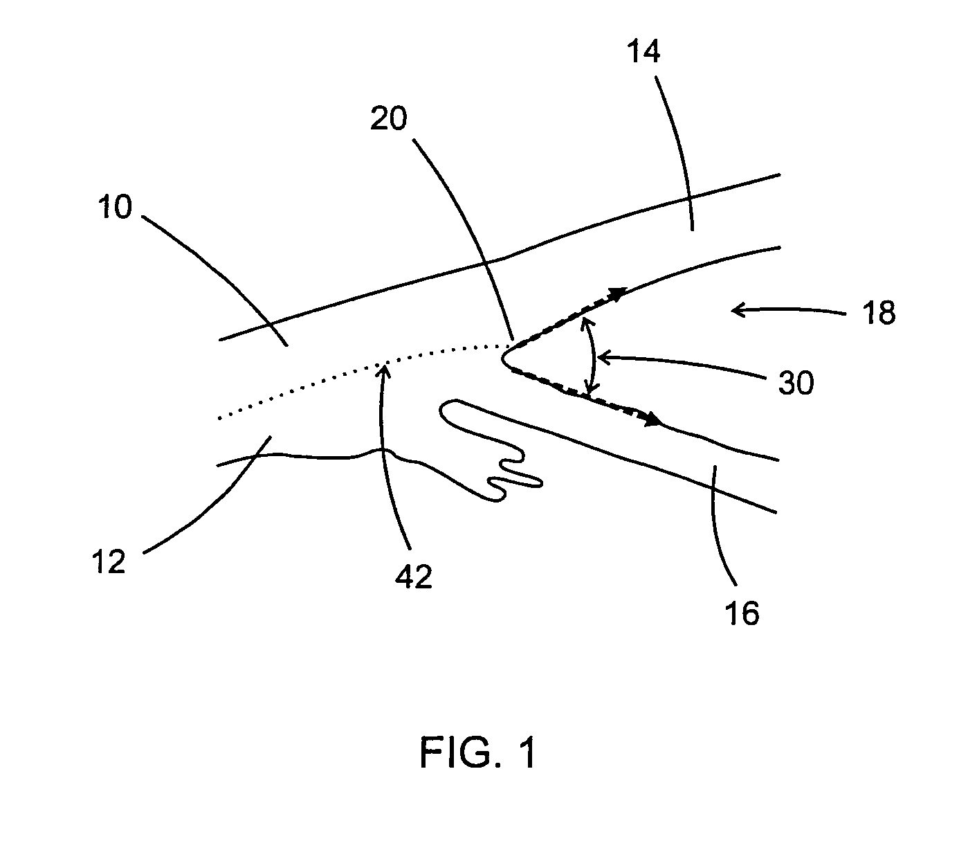
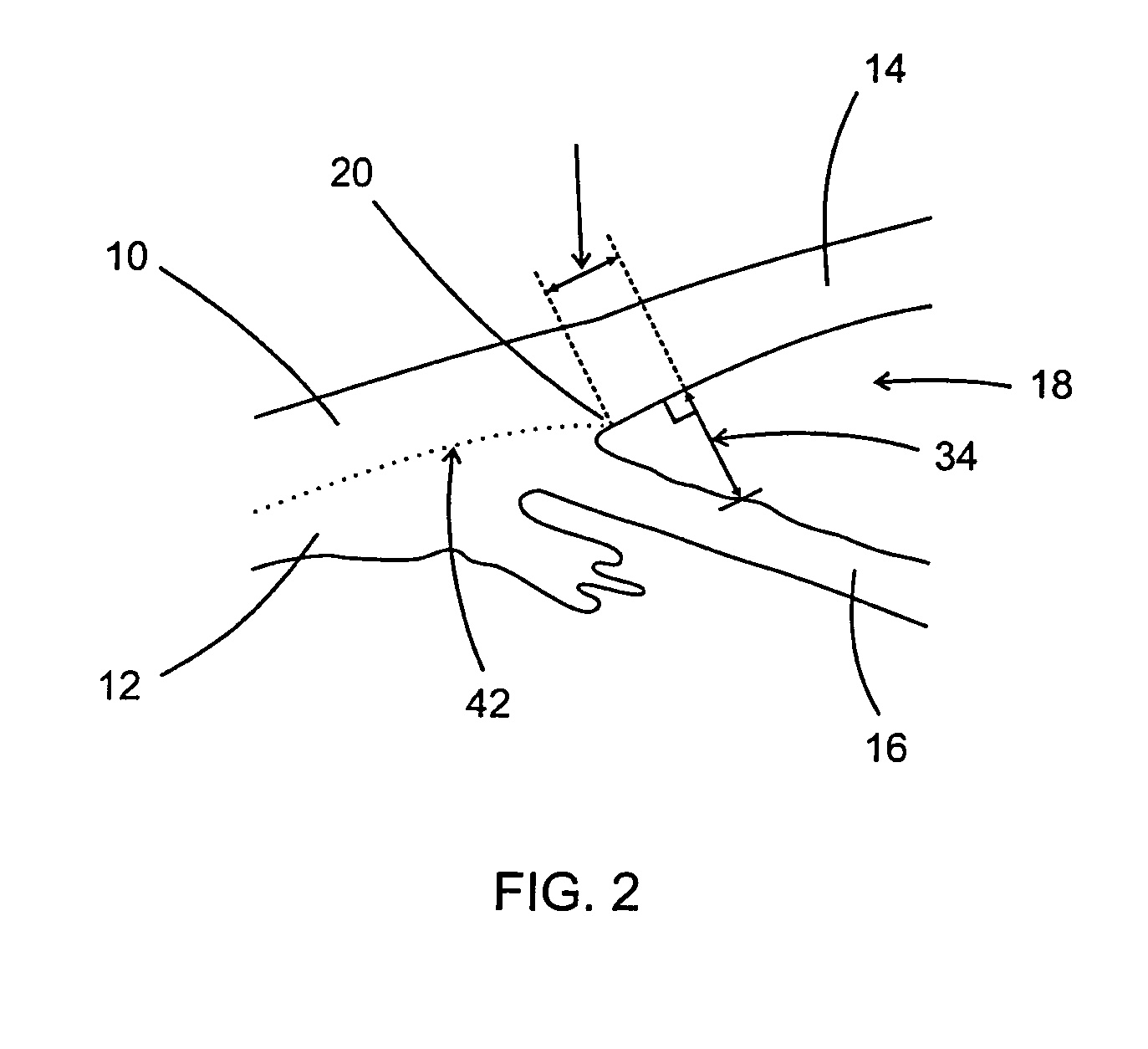
[0036]FIG. 1 illustrates key anatomical landmarks and measurements for examination of the anterior chamber of the eye. Shown are the locations of scleral tissue 10, uveal tissue 12, the scleral-uveal interface 42, the cornea 14, the iris 16, aqueous humor 18, the scleral spur 20, and the anterior chamber angle 30 is indicated. Anterior chamber angle 30 can be particularly useful in assessing the eye and diagnosing specific disease states or disorders, such as distinguishing between open-angle glaucoma, narrow-angle and closed-angle glaucoma. This distinction can be important in determining the appropriate course of any required therapy. Accurately determining the location of the scleral spur is important for proper evaluation of anterior chamber geometry. A proper evaluation of anterior chamber angles requires the location of scleral spur 20 to be known. The image requirements to identify the scleral spur:[0037]obtain image of the anterior chamber including the cornea and iris,[0038...


PUM


Abstract

A method of determining the location of the scleral spur, to allow measurements of the anterior chamber of the eye to facilitate diagnosis or treatment of the eye. The method utilizes ultrasonic biomicroscopy apparatus to image the anterior chamber and several millimeters of sclera and align indicators, or along the scleral-uveal interface and the corneal-aqueous interface. These indicators can be lines, dots or any shape or colors. The intersection of these indicators is used as an estimated location of the scleral spur for measuring anterior chamber angle, angle opening distance, or other aspects of the eye. The disclosure includes methods of locating the scleral spur, software for locating the scleral spur using computerized imaging apparatus, and systems including computerized imaging apparatus and software for locating the scleral spur.

Description

CROSS REFERENCES TO RELATED APPLICATIONS[0001]This patent application claims benefit from the earlier filed U.S. Provisional Application No. 61 / 854,726 filed Apr. 30, 2013, entitled “Ultrasonic Biomicroscopy (UBM) Methodology to Identify the Scleral Spur”, and is hereby incorporated into this application by reference as if fully set forth herein.BACKGROUND OF THE INVENTION[0002]1. Field of the Invention[0003]The present invention pertains to ophthalmic diagnosis and therapy, and more particularly, its apparatus, software, means, and methods for diagnosing and treating diseases of the eye. In particular, the present invention provides for improved evaluation of anterior chamber angle by accurately and repeatedly identifying the location of the scleral spur.[0004]The scleral spur is a subtle ridge of the sclera at the level of the limbus interposed between the posterior portion of Schlemm's canal and the anterior part of the ciliary body. The scleral spur is the structure to which som...

Claims


Application Information

Patent Timeline
IPC IPC(8): A61B3/00A61B3/14A61B3/117
CPCA61B3/117A61B3/102
Inventor PRAGER, THOMAS C.BURBA, THOMAS A.HARDTEN, DAVID R.
Owner ESI
Learn More