
Inter-organ spacer for use in endoscopic surgery

a technology for endoscopic surgery and interorgan spacers, which is applied in the field of interorgan spacers for use in endoscopic surgery, can solve the problems of degrading the endoscopic field, difficult hemostasis, and difficult maintenance of the endoscopic field, so as to reduce the risk of damage, and facilitate the operation.

Active Publication Date: 2015-02-03
NIPPON MEDICAL SCHOOL FOUND +1
View PDF28 Cites 0 Cited by
  • Summary
  • Abstract
  • Description
  • Claims
  • Application Information

AI Technical Summary

Benefits of technology

[0022]Use of the inter-organ spacer of the invention simultaneously solves various problems specific to endoscopic surgery. The advantages obtained include:
[0025](3) prevention of organ damage caused by the transfer of heat or oscillating waves and prevention of secondary damage when using an automatic suturing machine,
[0029]In endoscopic surgery, even a small amount of bleeding may interfere with the endoscopic field to impede smooth operation. However, when the inter-organ spacer of the invention is inserted, blood or exudates, such as bile or intestinal fluid, can be immediately absorbed into the liquid holding means, preventing it from diffusing to the surroundings.
[0031]In endoscopic surgery, the target organ or tissue is subjected to exclusion through the use of instruments, such as forceps, by remote control. This is because the target organ or tissue cannot be protectively pressed (subjected to exclusion) with the hand or fingers. In this process, the force may often be concentrated at one point, causing damage to the organ or tissue. The inter-organ spacer of the invention may be placed between the forceps and the organ or tissue to dissipate the force so that it is no longer concentrated, reducing the risk of damage.

Problems solved by technology

the effect of magnification by laparoscope, which makes possible delicate techniques with less bleeding.
However, endoscopic surgery has the following problems.
Once bleeding occurs, the endoscopic field is degraded, and hemostasis is more difficult than in laparotomy.
Therefore, when exudates and blood are sucked during surgery, abdominal gas (CO2) is simultaneously sucked, so that the endoscopic field can be difficult to maintain, which makes suction more difficult than in laparotomy.
In two-dimensional monitoring, it is difficult to monitor organs in the deep field, which creates the risk of damage to organs.
Organs cannot be directly touched by hand and fingers, and operations are performed using hard forceps, which makes it difficult to protectively treat organs.
These problems are common to all endoscopic operations of abdominal organs, which include operations in the field of not only digestive surgery but also gynecology and urology.
As described above, the problems with endoscopic surgery are not significant for laparotomy with a long history, and therefore, there is no medical device capable of solving the problems found in the medical devices for laparotomy.
In addition, at present, these instruments are used only in very restricted situations.
In endoscopic surgery, however, it is difficult to frequently put gauze in and out through a trocar, and the replacement with new gauze is not easy, in contrast to laparotomy.
However, gauze is thin and relatively weak and therefore creates the risk of damage to an organ from the tip of a suction tube in the process of suction through the suction tube.
In addition, gauze may adhere to the organ when dried, and the adhering gauze may cause damage to the organ when peeled off.
It is also difficult to appropriately fold gauze in the abdominal cavity, which causes the disadvantage that it is difficult to protectively wrap or press an organ as in laparotomy.

Method used


Image

  • Inter-organ spacer for use in endoscopic surgery
  • Inter-organ spacer for use in endoscopic surgery
  • Inter-organ spacer for use in endoscopic surgery

Examples


example 1

Evaluation of Liquid Absorbency

[0098]This example and each example described below were performed using three porous soft materials (hereinafter referred to as sample set A): a polyurethane material with low liquid releasing ability (Toughponge (Hogy Medical Co., Ltd.)); a polyurethane material with high liquid releasing ability for abdominal surgery; and a polyvinyl alcohol material (Retractor Pad (Heiwa Iryo Kikai Co., Ltd.)). Another sample set used includes six materials (hereinafter referred to as sample set B): the polyurethane material with low liquid release ability (Toughponge (Hogy Medical Co., Ltd.), hereinafter also referred to as sample A); four different polyurethane materials with high liquid releasing ability (hereinafter also referred to as samples B to E); and a polyethylene material (hereinafter also referred to as sample F). Table 2 shows the principal properties of each porous soft material used in sample sets A and B. The elongation in Table 2 was measured acco...

example 2

Evaluation of Liquid Holding Ability

[0108]The weight (Wd in units of g) of the sample (in the shape of a rectangular parallelepiped of 20 mm×20 mm×80 mm) was measured before it was allowed to absorb artificial blood. After the sample was allowed to absorb a sufficient amount of artificial blood (Synthetic Blood Reagent Mix (Johnson, Moen & Co.)), its weight (Ww in units of g) was measured. Subsequently, the sample was placed on a polyvinyl chloride plate, and the plate was allowed to stand for 3 minutes, while being tilted at 45°. Thereafter, the weight (W3 in units of g) of the sample was measured.

[0109]The liquid holding ability was calculated from the following formula: (W3−Wd)÷(Ww−Wd)×100(%).

[0110]The results on sample sets A and B are shown in Tables 5 and 6, respectively. Three samples were prepared for each material, and the average of the liquid holding abilities (%) of the respective samples was calculated. The higher value indicates the higher liquid holding ability.

[0111]...

example 3

Evaluation of Liquid Releasing Ability

[0113]The weight (Wd in units of g) of the sample (in the shape of a rectangular parallelepiped of 20 mm×50 mm×50 mm) was measured before it was allowed to absorb artificial blood. After the sample was allowed to absorb a sufficient amount of artificial blood (Synthetic Blood Reagent Mix (Johnson, Moen & Co.)), it was placed on a plastic plate, and its weight (Ww in units of g) was measured. The artificial blood was sucked under a reduced pressure of −600 to −650 mmHg (−80 to −85 kPa) for 1 minute, while the tip of a Yanker suction tube was held to the center of the sample and pushed in. After the suction, the weight (Ws in units of g) of the sample being placed on the plastic plate was measured. The liquid releasing ability was calculated from the following formula: (Ww−Ws)÷(Ww−Wd)×100(%).

[0114]The results on sample sets A and B are shown in Tables 7 and 8, respectively. Three samples were prepared for each material, and the average of the liqu...


PUM


Abstract

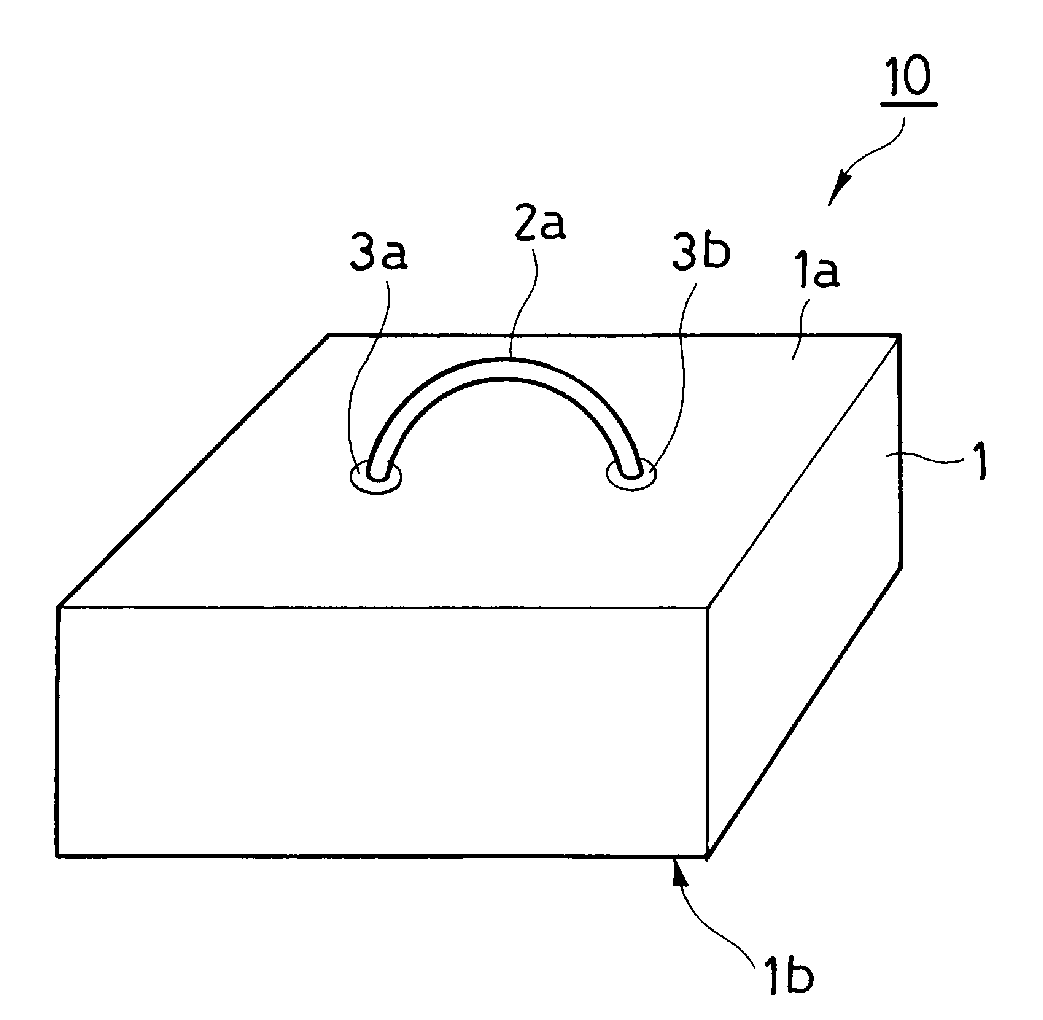
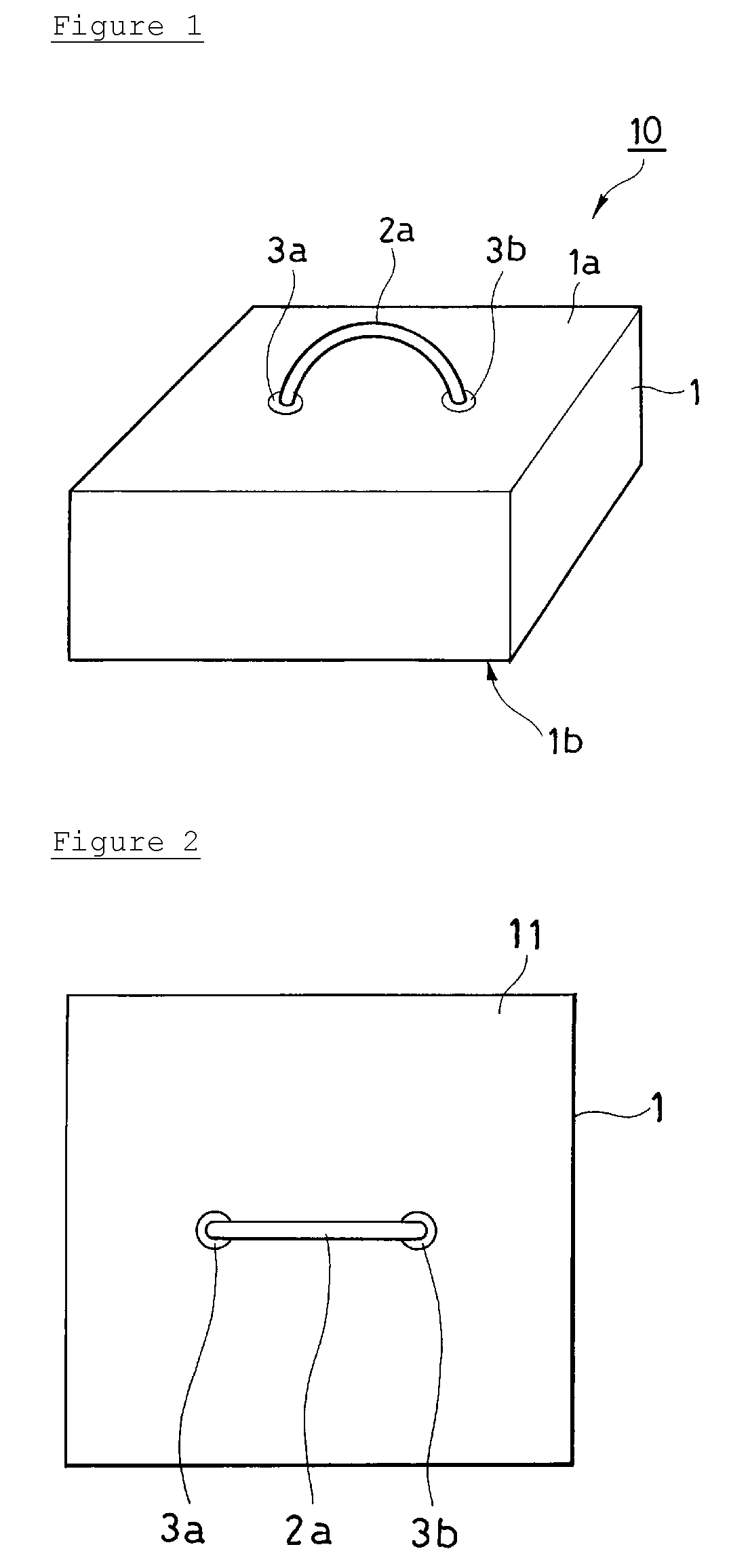
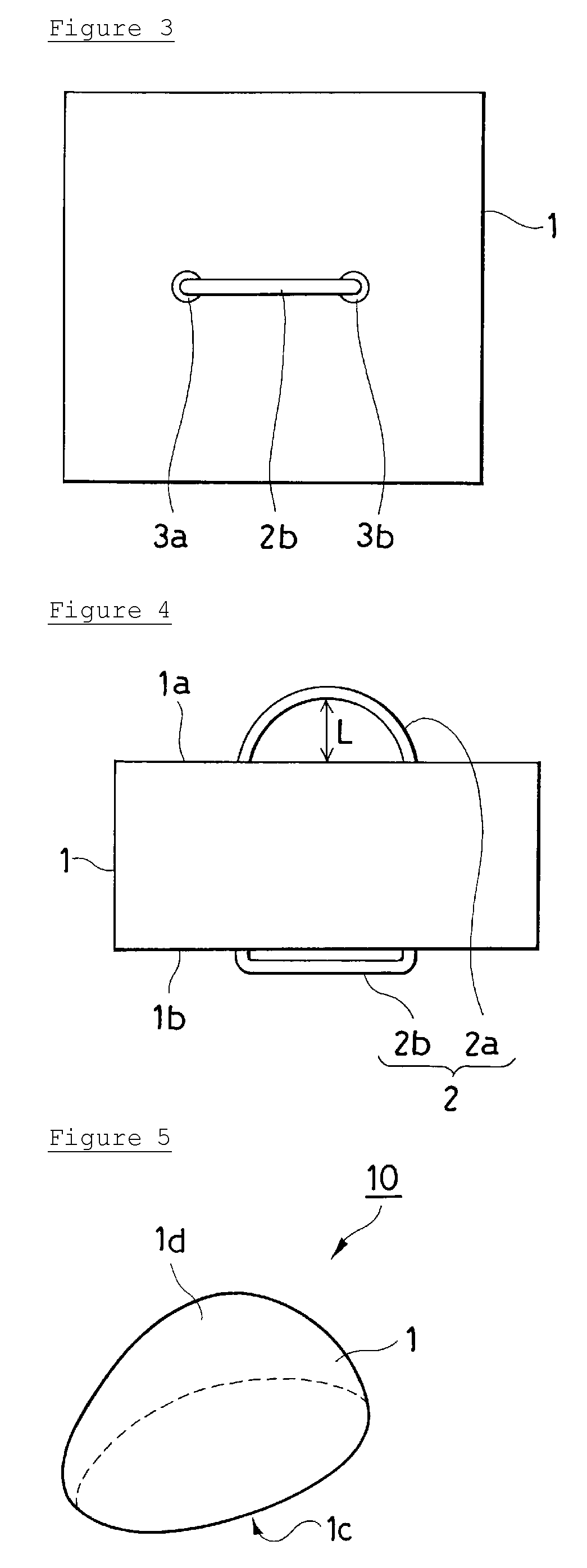
The invention provides a surgical device for endoscopic surgery having the function of temporarily storing liquid (a first function), the function of keeping a physical space for surgery and a sufficient endoscopic field (a second function), and the function of protecting organs (a third function), and an insertion device for inserting the surgical device into a body.[Solving Means]The surgical device for endoscopic surgery is an inter-organ spacer (10) including liquid holding means (1) formed of a porous soft material having the ability to temporarily store liquid and the ability to retain its shape. The insertion device for inserting the inter-organ spacer into a body includes a tubular holder capable of holding the inter-organ spacer for endoscopic surgery.

Description

CROSS REFERENCE TO RELATED APPLICATIONS[0001]This application is a national phase filing under 35 U.S.C. §371 of PCT International Application PCT / JP2008 / 073859, filed Dec. 26, 2008, and published under PCT Article 21(2) in Japanese as WO 2009 / 084688 A1 on Jul. 9, 2009. PCT / JP2008 / 073859 claims priority from Japanese application No. 2007-340837 filed on Dec. 28, 2007. The entire contents of each of the prior applications are incorporated herein by reference.TECHNICAL FIELD[0002]The present invention relates to an inter-organ spacer for use in endoscopic surgery and an insertion device for inserting the spacer into a body.BACKGROUND ART[0003]Endoscopic surgery includes thoracic or abdominal surgery performed under the monitoring of images from an endoscope (e.g., thoracoscope or laparoscope) inserted in a thoracic or abdominal cavity. In laparoscopic surgery, for example, unlike traditional abdominal surgery (laparotomy), incisions in the abdomen are not large, and operations are per...

Claims


Application Information

Patent Timeline
Patent Type & Authority Patents(United States)
IPC IPC(8): A61F2/00A61F13/00A61F13/20A61B17/00A61B17/02A61B19/00
CPCA61B17/02A61B17/0218A61B2019/4878A61B2017/00898A61B2090/0815
Inventor NAKAMURA, YOSHIHARUMATSUMOTO, SATOSHIINOUE, TOSHIKI
Owner NIPPON MEDICAL SCHOOL FOUND
Learn More