System and method for quantitative molecular breast imaging

a molecular breast and molecular imaging technology, applied in the field of molecular breast imaging systems and methods, can solve the problems of reducing the usefulness of this procedure, not being widely used as a screening technique, and the cost of mri is currently approximately 20 times more, so as to achieve accurate size determination

Active Publication Date: 2014-12-30
MAYO FOUND FOR MEDICAL EDUCATION & RES
View PDF15 Cites 5 Cited by
  • Summary
  • Abstract
  • Description
  • Claims
  • Application Information

AI Technical Summary

Benefits of technology

[0011]The present invention provides a system and method for performing quantitative tumor analysis using information acquired with a dual-headed molecular breast imaging system. Specifically, the present invention provides systems and method to utilize the information available in planar dedicated breast imaging to provide previously unavailable information sets to aid in the diagnosis and biopsy of the site. In particular, the present invention provides a method for accurately determining the size, depth to the collimator, and relative tracer uptake of a tumor.

Problems solved by technology

However, since bilateral breast MRI is currently approximately 20 times more expensive than mammography, it is not in widespread use as a screening technique.
The PEM technology offers excellent resolution; however, the currently available radiotracer (F-18 Fluoro deoxyglucose) requires that a patient fast overnight, the patient must have low blood levels (this is often a problem for diabetics), and after injection, the patient must wait 1-2 hours for optimum uptake of F-18FDG in the tumor.
The high cost of these PET procedures coupled with the long patient preparation time reduces the usefulness of this procedure and makes it difficult to employ for routine breast evaluation.
However, these results only hold for large tumors and several studies have shown that the sensitivity falls significantly with tumor size.
This limitation is particularly important in light of the finding that up to a third of breast cancers detected by screening mammography are smaller than 10 mm.
The failure of conventional scintimammography to meet this limit led to its abandonment as a useful technique in the United States.
Despite this improved percentage of success, the failure to identify lesions of any size can have significant consequences.

Method used


Image

  • System and method for quantitative molecular breast imaging
  • System and method for quantitative molecular breast imaging
  • System and method for quantitative molecular breast imaging

Examples


Embodiment Construction

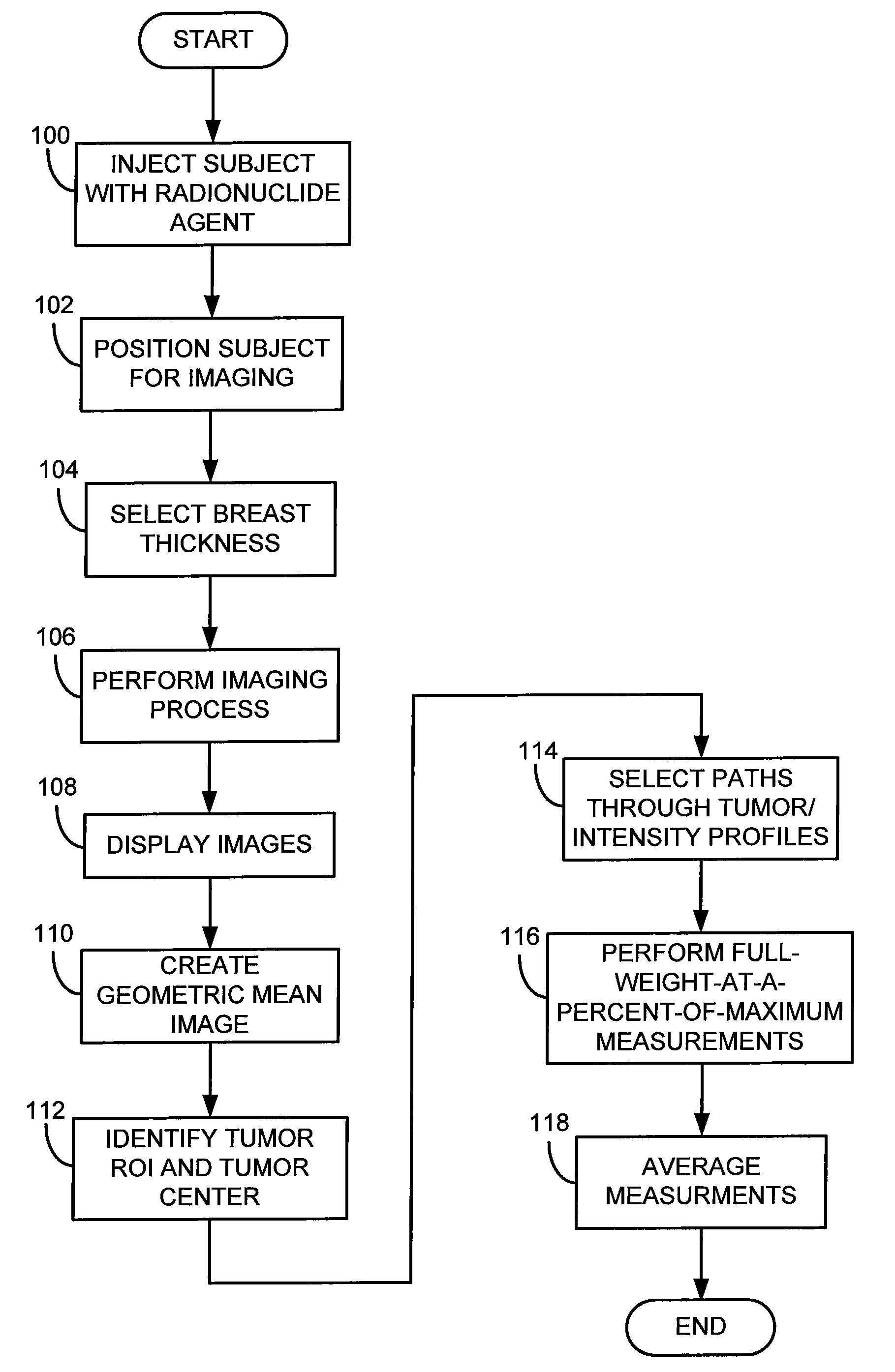
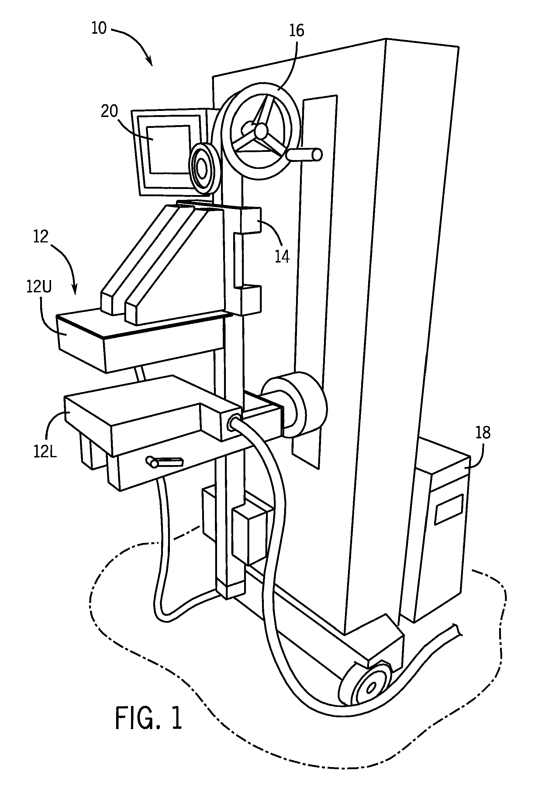
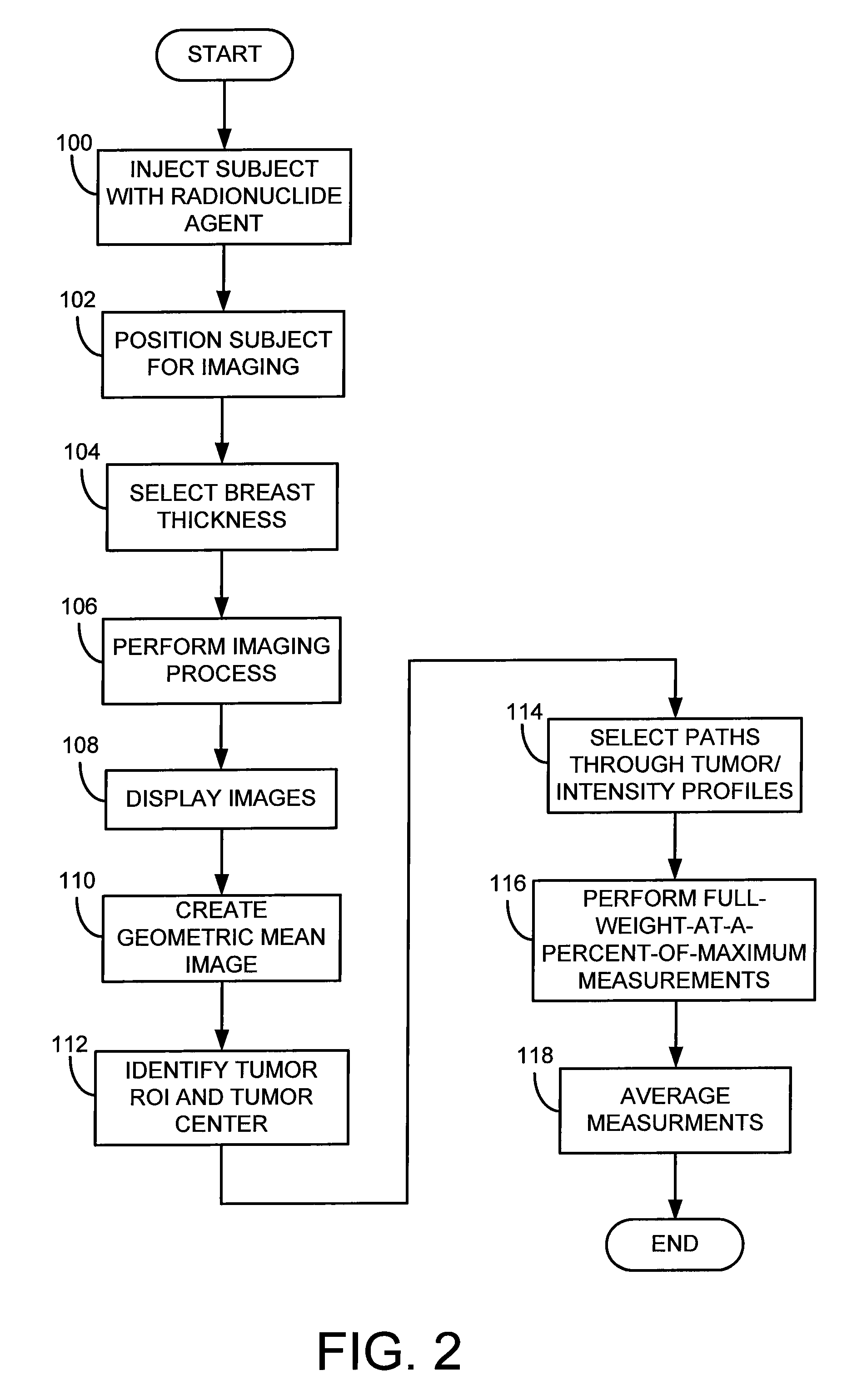
[0019]Referring to FIG. 1, a molecular breast imaging (MBI) system 10 includes two opposing cadmium zinc telluride (CZT) detectors (detector heads) 12. In particular, the detector heads 12 include an upper detector head 12U and a lower detector head 12L. Each detector head 12U, 12L is, for example, 20 cm by 16 cm in size and mounted on a modified upright type mammographic gantry 14. In accordance with one embodiment, the detector heads 12 are LumaGEM 3200S high-performance, solid-state cameras from Gamma Medica having a pixel size of 1.6 mm. LumaGEM is a trademark of Gamma Medica, Inc. Corporation of California.

[0020]The relative position of the detector heads 12 can be adjusted using a user control 16. Specifically, the detector head assemblies 12 are, preferably, designed to serve as a compression mechanism. Accordingly, this system configuration reduces the maximum distance between any lesion in the breast and either detector head 12 to one-half of the total breast thickness, pot...


PUM

PropertyMeasurementUnit
sizeaaaaaaaaaa
sizeaaaaaaaaaa
diameteraaaaaaaaaa

Abstract

A system and method for performing quantitative lesion analysis in molecular breast imaging (MBI) using the opposing images of a slightly compressed breast that are obtained from the dual-head gamma camera. The method uses the shape of the pixel intensity profiles through each tumor to determine tumor diameter. Also, the method uses a thickness of the compressed breast and the attenuation of gamma rays in soft tissue to determine the depth of the tumor from the collimator face of the detector head. Further still, the method uses the measured tumor diameter and measurements of counts in the tumor and background breast region to determine relative radiotracer uptake or tumor-to-background ratio (T / B ratio).

Description

CROSS-REFERENCE TO RELATED APPLICATIONS[0001]The present application is based on U.S. Provisional Patent Application Ser. No. 60 / 869,419, entitled “Quantitative Molecular Breast Imaging Using Dual Head Gamma Cameras,” filed Dec. 11, 2006, and claims the benefit thereof.STATEMENT REGARDING FEDERALLY SPONSORED RESEARCH[0002]This invention was made with government support under Grant No. CA110162 awarded by the National Cancer Institute. The United States Government has certain rights in this invention.FIELD OF THE INVENTION[0003]The invention relates to a system and method for obtaining quantitative information regarding breast images acquired using gamma cameras.BACKGROUND OF THE INVENTION[0004]Screening mammography has been the gold standard for breast cancer detection for over 30 years, and is the only available screening method proven to reduce breast cancer mortality. However, the sensitivity of screening mammography varies considerably. The most important factor in the failure o...

Claims


Application Information

Patent Timeline
Patent Type & Authority Patents(United States)
IPC IPC(8): G06K9/00G01T1/164G01N23/00A61B5/05A61B6/00
CPCG01T1/1644A61B6/4258
Inventor O'CONNOR, MICHAEL K.HRUSKA, CARRIE B.
Owner MAYO FOUND FOR MEDICAL EDUCATION & RES
Why Eureka
Try Eureka