Eureka AIR delivers breakthrough ideas for toughest innovation challenges, trusted by R&D personnel around the world.

3-dimensional cardiac fibroblast derived extracellular matrix

a fibroblast and extracellular matrix technology, applied in the field of 3-dimensional cardiac fibroblast derived extracellular matrix, can solve the problems of inability to demonstrate robust cardiac regeneration, lack of unique structural characteristics, and inability to engraft large-scale cells or functional cardiomyocyte regeneration in infarcted hearts, so as to facilitate the delivery of seeded cells and move flexibly.

Active Publication Date: 2014-08-12
WISCONSIN ALUMNI RES FOUND
View PDF6 Cites 8 Cited by
  • Summary
  • Abstract
  • Description
  • Claims
  • Application Information

AI Technical Summary

Benefits of technology

[0011]This application discloses an epicardial patch for facilitating the delivery of cells to myocardial tissue, as well the isolated cardiac extracellular matrix that makes up the patch and methods of making and using the same. Advantageously, the isolated cardiac extracellular matrix is truly 3-dimensional, is myocardial in origin and composition, adheres well to the wall of the heart without the need for glue or sutures, moves flexibly with the heart, and successfully facilitates the delivery of seeded cells into myocardial tissue.

Problems solved by technology

This slow cardiomyocyte turnover is important for maintaining cardiac function from birth to old age; however, resident cardiac progenitor cells have shown little capacity for robust cardiac regeneration following myocardial injury such as that caused by a myocardial infarction (see Bolli, P. and H. W. Chaudhry, Molecular physiology of cardiac regeneration.
A major obstacle in developing cell-based regenerative strategies is the need to successfully transfer a sufficient number of therapeutic cells to the target location (Karp, J. M. and G. S. Leng Teo, Mesenchymal stem cell homing: the devil is in the details.
For example, in the infarcted heart, transfer of therapeutic cells is challenged not only by the motion of the heart but also by its heightened electrical and structural instability (Fernandes, S., et al., Autologous myoblast transplantation after myocardial infarction increases the inducibility of ventricular arrhythmias.
However, to date, no study has reported the large scale engraftment of cells or functional cardiomyocyte regeneration in an infarcted heart.
Notably, none of these patches are made from myocardial tissue, nor do any of them exhibit the unique structural characteristics of cardiac ECM.
Accordingly, transplantation of cells across different tissue types could be problematic (see Badylak, S. F., D. O. Freytes, and T. W. Gilbert, Extracellular matrix as a biological scaffold material: Structure and function.
In addition to not being of myocardial origin, biomaterials currently under investigation for use in cardiac cell transfer patches have other notable limitations.
For example, a frequent issue with the use of synthetic or decellularized tissues in patches for therapeutic cell delivery to the myocardium is the inability of the patch to physically adhere to the epicardial surface.
If a patch's compliance does not match that of the ventricle and does not move with the beating heart, gaps may form under the surface, reducing cell transfer.

Method used


Image

  • 3-dimensional cardiac fibroblast derived extracellular matrix
  • 3-dimensional cardiac fibroblast derived extracellular matrix
  • 3-dimensional cardiac fibroblast derived extracellular matrix

Examples


example 1

Production and Characterization of Cardiac Derived 3-Dimensional Extracellular Matrix

[0061]In this Example, we generated a 3D-ECM using cardiac fibroblasts that is measured by confocal microscopy to be between 30-150+ μm thick. Additionally, we devised multiple ways to remove the fibroblasts (decellularization), which allow for the study of the effect of cellular debris on stem cell attachment, proliferation and differentiation. Controlling (varying) the cell debris content is important because cellular debris is highly prevalent in the early phases of cardiac healing, and has largely been overlooked as a factor impacting therapeutic cell adhesion and differentiation. This new method of generating cardiac specific extracellular matrix will facilitate in vitro studies of stem cell interactions with a cardiac specific matrix.

[0062]An important first step in studying the CF 3D-ECM is carrying out detailed analyses of its composition and structure. This was done using bottom-up 2D-(stro...

example 2

Dynamic Adhesion of Mesenchymal Stem Cells to Cardiac Fibroblast 3-Dimensional Extracellular Matrix

[0111]Therapeutic cell adhesion and engraftment are essential to the success of cell-based therapy. Current techniques involving circulatory infusion (systemic or intracoronary) have been used in large scale clinical trials with mixed results, with extremely low reported retention rates for both systemic and intracoronary infusion. Intramyocardial injection (either epicardial or endocardial) tends to have greater cell retention rates than circulatory delivery modes and has the ability to deliver cells with limited extravasation potential, such as skeletal myoblasts. To date, studies quantifying cell retention in vivo have been difficult and expensive to perform.

[0112]To address this issue, we utilized an oscillatory dynamic adhesion assay developed by others to test the adhesion of BMSC to our CF 3D-ECM generated with AH or PAA and to cardiac fibroblasts (non-decellularized). We also d...

example 3

Cardiac Fibroblast Patches: Successful Adhesion of Epicardial Patch to Epicardium

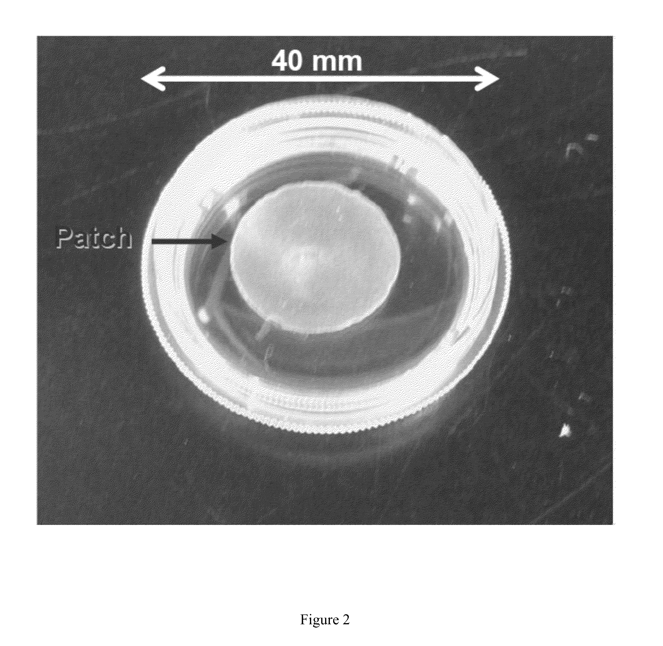
[0138]Epicardial patches have emerged as a promising mode of therapeutic cell delivery for cardiac regeneration. One issue with using synthetic or decellularized tissues as patch materials is the inability of the patch to physically adhere to the surface epicardium, often requiring the use of glue or sutures to hold the patch to the heart. If the patch does not maintain firm contact with the surface of the heart, the ability of the cells to transfer is decreased significantly. We demonstrate in this Example that CF 3D-ECM adheres well to the surface of a heart, creating a unique cardiac based platform for therapeutic cell transfer. This work represents collaboration with the laboratory of Professor Brenda Ogle of the Stem cell and Regenerative Medicine Center at the University of Wisconsin-Madison. Nicholas Kouris of Professor Ogle's laboratory provided human mesenchymal stem cells and carried out the C...


PUM

PropertyMeasurementUnit
thicknessaaaaaaaaaa
thicknessaaaaaaaaaa
thicknessaaaaaaaaaa

Abstract

A bioscaffold made from an isolated cardiac fibroblast-derived 3-dimensional extracellular matrix (ECM) is disclosed. The bioscaffold can be used as an epicardial patch for the delivery of therapeutic cells into myocardial tissue. Methods of making the 3-dimensional extracellular matrix using cultured cardiac fibroblasts are also disclosed.

Description

CROSS-REFERENCE TO RELATED APPLICATIONS[0001]This application claims the benefit of U.S. provisional Application No. 61 / 575,658 filed on Aug. 25, 2011, which is incorporated by reference herein in its entirety.STATEMENT REGARDING FEDERALLY SPONSORED RESEARCH OR DEVELOPMENT[0002]This invention was made with government support under HL092477 awarded by the National Institutes of Health. The government has certain rights in the invention.BACKGROUND[0003]The human heart continuously undergoes slow cellular turnover through apoptosis of cardiomyocytes and proliferation of cardiac progenitor cells at a rate of approximately 1% turnover a year (see Bergmann, O., et al., Evidence for cardiomyocyte renewal in humans. Science, 2009. 324(5923): p. 98-102). This slow cardiomyocyte turnover is important for maintaining cardiac function from birth to old age; however, resident cardiac progenitor cells have shown little capacity for robust cardiac regeneration following myocardial injury such as t...

Claims


Application Information

Patent Timeline
Patent Type & Authority Patents(United States)
IPC IPC(8): A61P9/00A61K35/48A61K47/42C12P21/00A61K35/36A61K35/28A61K35/34A61K35/00A61K9/00A61K35/32A61K35/33
CPCA61L27/54A61K35/33A61L27/3834A61L27/3633A61K35/34C12N5/0657A61K35/28A61L2300/414A61L2430/20A61K38/00A61P9/00A61K2300/00A61L27/3687C12N5/0663C12N2513/00C12N2533/90
Inventor SCHMUCK, ERIC G.SAUPE, KURT W.
Owner WISCONSIN ALUMNI RES FOUND
Why Eureka
Eureka Blog
Learn More